Copyright
©The Author(s) 2024.
World J Gastrointest Oncol. May 15, 2024; 16(5): 2253-2260
Published online May 15, 2024. doi: 10.4251/wjgo.v16.i5.2253
Published online May 15, 2024. doi: 10.4251/wjgo.v16.i5.2253
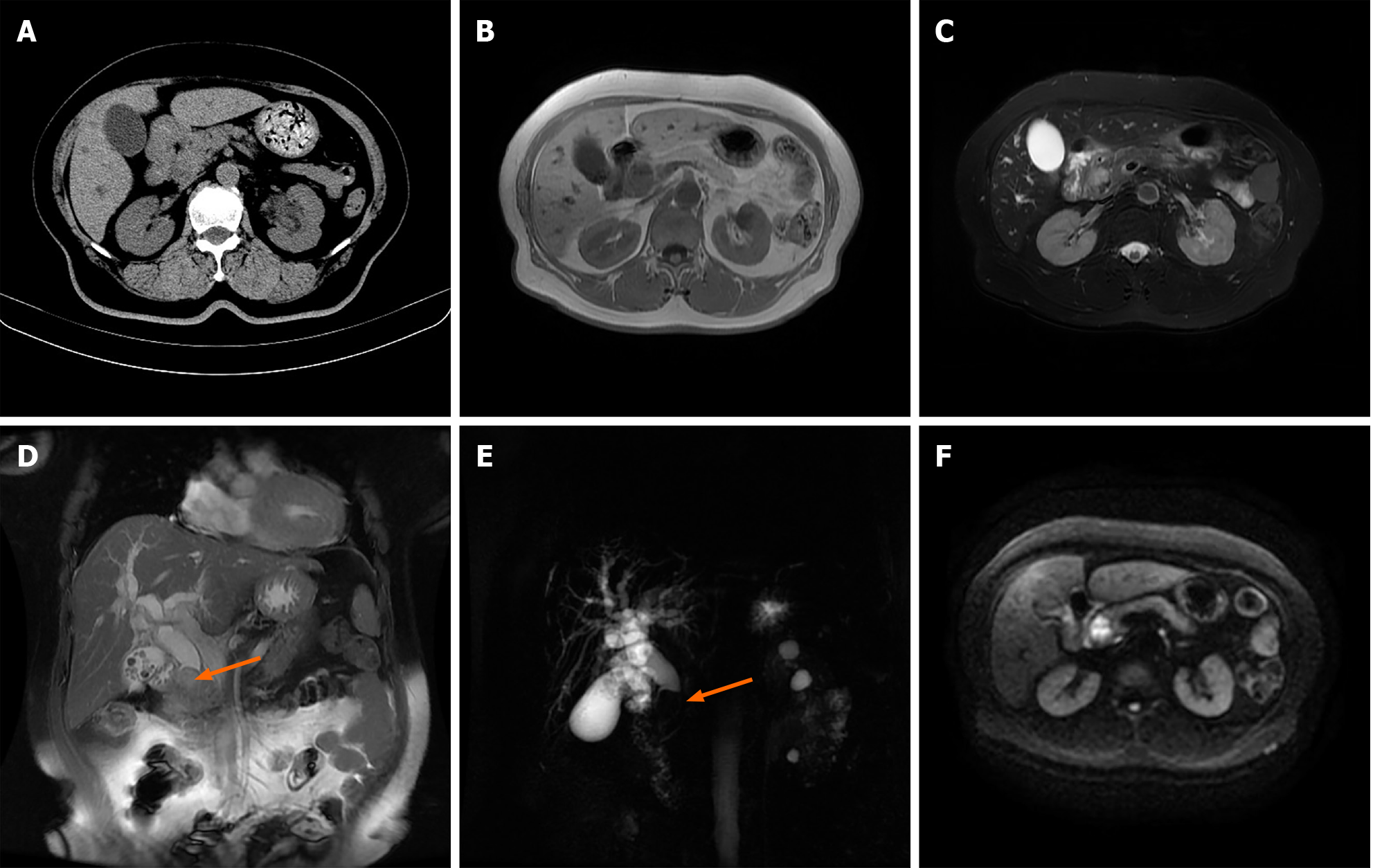
Figure 1 Imaging revealed calculous cholecystitis, intrahepatic and extrahepatic bile duct dilation with extrahepatic bile duct calculi, and a space-occupying lesion of the distal common bile duct.
A: Computed tomography showed calculous cholecystitis, and a soft tissue density shadow at the distal common bile duct (CBD) with intrahepatic and extrahepatic bile duct dilatation; B-F: Magnetic resonance imaging by the (B) T1-weighted image, (C) T2-weighted image, (D) coronal plane sequence, (E) magnetic resonance cholangiopancreatography and (F) diffusion-weighted imaging revealed calculous cholecystitis, intrahepatic and extrahepatic bile duct dilation with extrahepatic bile duct calculi, and a soft tissue mass signal at the distal CBD with limited diffusion.
- Citation: Zheng LP, Shen WY, Hu CD, Wang CH, Chen XJ, Wang J, Shen YY. Undifferentiated high-grade pleomorphic sarcoma of the common bile duct: A case report and review of literature. World J Gastrointest Oncol 2024; 16(5): 2253-2260
- URL: https://www.wjgnet.com/1948-5204/full/v16/i5/2253.htm
- DOI: https://dx.doi.org/10.4251/wjgo.v16.i5.2253


