Published online Apr 15, 2024. doi: 10.4251/wjgo.v16.i4.1256
Peer-review started: November 5, 2023
First decision: December 20, 2023
Revised: December 27, 2023
Accepted: February 1, 2024
Article in press: February 1, 2024
Published online: April 15, 2024
Processing time: 157 Days and 22 Hours
One of the primary reasons for the dismal survival rates in pancreatic ductal adenocarcinoma (PDAC) is that most patients are usually diagnosed at late stages. There is an urgent unmet clinical need to identify and develop diagnostic methods that could precisely detect PDAC at its earliest stages.
To evaluate the potential value of radiomics analysis in the differentiation of early-stage PDAC from late-stage PDAC.
A total of 71 patients with pathologically proved PDAC based on surgical resection who underwent contrast-enhanced computed tomography (CT) within 30 d prior to surgery were included in the study. Tumor staging was performed in accordance with the 8th edition of the American Joint Committee on Cancer staging system. Radiomics features were extracted from the region of interest (ROI) for each patient using Analysis Kit software. The most important and predictive radiomics features were selected using Mann-Whitney U test, univar
A total of 792 radiomics features (396 from late arterial phase and 396 from portal venous phase) were extracted from the ROI for each patient using Analysis Kit software. Nine most important and predictive features were selected using Mann-Whitney U test, univariate logistic regression analysis, and MRMR method. RF method was used to construct the radiomics model with the nine most predictive radiomics features, which showed a high discriminative ability with 97.7% accuracy, 97.6% sensitivity, 97.8% specificity, 98.4% positive predictive value, and 96.8% negative predictive value. The radiomics model was proved to be robust and reproducible using 10-times LGOCV method with an average area under the curve of 0.75 by the average performance of the 10 newly built models.
The radiomics model based on CT could serve as a promising non-invasive method in differential diagnosis between early and late stage PDAC.
Core Tip: Pancreatic ductal adenocarcinoma (PDAC) remains the deadliest of the common cancers, with little change in patient survival in the past several decades. One of the biggest challenges of the management of PDAC that physicians often encounter is that the early detection in high-risk individuals and the early diagnosis of patients with suspected symptoms. Precise staging of PDAC is vital not only in making treatment decisions, but also in evaluating prognosis. Radiomics, the generation of minable high throughput data through conversion of digital computed tomography (CT) or magnetic resonance imaging, allows obtaining additional insight into pancreatic tissue heterogeneity. The aim of our study was to investigate a radiomics approach for potential differentiation of early- from late-stage PDAC. In conclusion, our study demonstrated that the radiomics model based on CT could serve as a promising non-invasive method in differential diagnosis between early and late stage PDAC. Large-scale prospective cohort studies, preferably multi-center, to validate the potential value of the radiomics diagnostic approach in differentiating early from late stage PDAC are in order.
- Citation: Ren S, Qian LC, Cao YY, Daniels MJ, Song LN, Tian Y, Wang ZQ. Computed tomography-based radiomics diagnostic approach for differential diagnosis between early- and late-stage pancreatic ductal adenocarcinoma. World J Gastrointest Oncol 2024; 16(4): 1256-1267
- URL: https://www.wjgnet.com/1948-5204/full/v16/i4/1256.htm
- DOI: https://dx.doi.org/10.4251/wjgo.v16.i4.1256
Pancreatic cancer remains one of the most dismal types of cancers worldwide, characterized by a poor prognosis and a low 5-year survival rate[1]. Pancreatic ductal adenocarcinoma (PDAC) constitutes more than 90% of all pancreatic cancer cases. PDAC is often diagnosed as advanced due to the anatomical structure of the deep retroperitoneal layer of the pancreas, lack of typical symptoms and effective screening methods[2]. PDAC is expected to surpass both breast and colorectal cancer as the second leading cause of cancer related deaths by year 2030[3].
One of the biggest challenges of the management of PDAC that physicians often encounter is that the early detection in high-risk individuals and the early diagnosis of patients with suspected symptoms. Two biomarkers including carbohydrate antigen 19-9 (CA 19-9) and carcinoembryonic antigen are recommended for screening PDAC in clinical practice[4,5]. CA 19-9 facilitates surgical decision and the detection of post-operative tumor recurrence; however, the utility of CA 19-9 is limited since 10% of patients do not secrete CA 19-9[6]. CA 19-9 levels are not found to be specific or sensitive enough for reliable detection of PDAC[5].
Multi-detector computed tomography (MDCT) is the most widely available and important pre-operative examination for patients with suspected PDAC[7]. MDCT provides good spatial resolution with wide anatomic coverage, which allows comprehensive evaluation of local and distant disease in a single session[8]. Notably, MDCT is proved to be a useful tool in assessment of vascular involvement, which is one of the deciding factors of tumor resectability[9,10]. The American Joint Committee on Cancer (AJCC) TNM staging criteria has been developed to characterize local and systemic spread PDAC. Precise staging of PDAC is vital not only in making treatment decisions, but also in evaluating prognoses[11]. The diagnosis and clinical staging of PDAC are established using CT and/or magnetic resonance imaging (MRI), or endoscopic ultrasound-guided fine needle aspiration[12].
Over the past few years, radiomics has facilitated the development of processes for the conversion of digital images into mineable data and further analysis of the data for decision support. In clinical practice, the specific biopsy sample can provide histological information of tumors but may not reflect the full extent of the tumor phenotype due to sampling error[12]. Radiomics is a promising useful tool that assesses tissue gray-level intensity and pixel position in digital CT or MRI images and permits quantification of tumor spatial heterogeneity, which can preoperatively predict histological grade and guide clinical decision-making. Radiomics has been used for pathologic classification and TNM staging in different tumors and initial results are encouraging[13-15]. Recently, a well-written and insightful article by Xu et al[16] investigated the potential value of a circular RNA (circRNA)-based biomarker panel in distinguishing between early (stage I/II)- and late-stage (stage III/IV) PDAC, which appeared in the October 2023 issue of Gastroenterology. In our study, we also classified all PDAC into early (stage I/II)- and late-stage (stage III/IV), and investigated the potential value of radiomics model in distinguishing between early- and late- stage PDAC, which is critical for making strategic management decision and predicting prognoses.
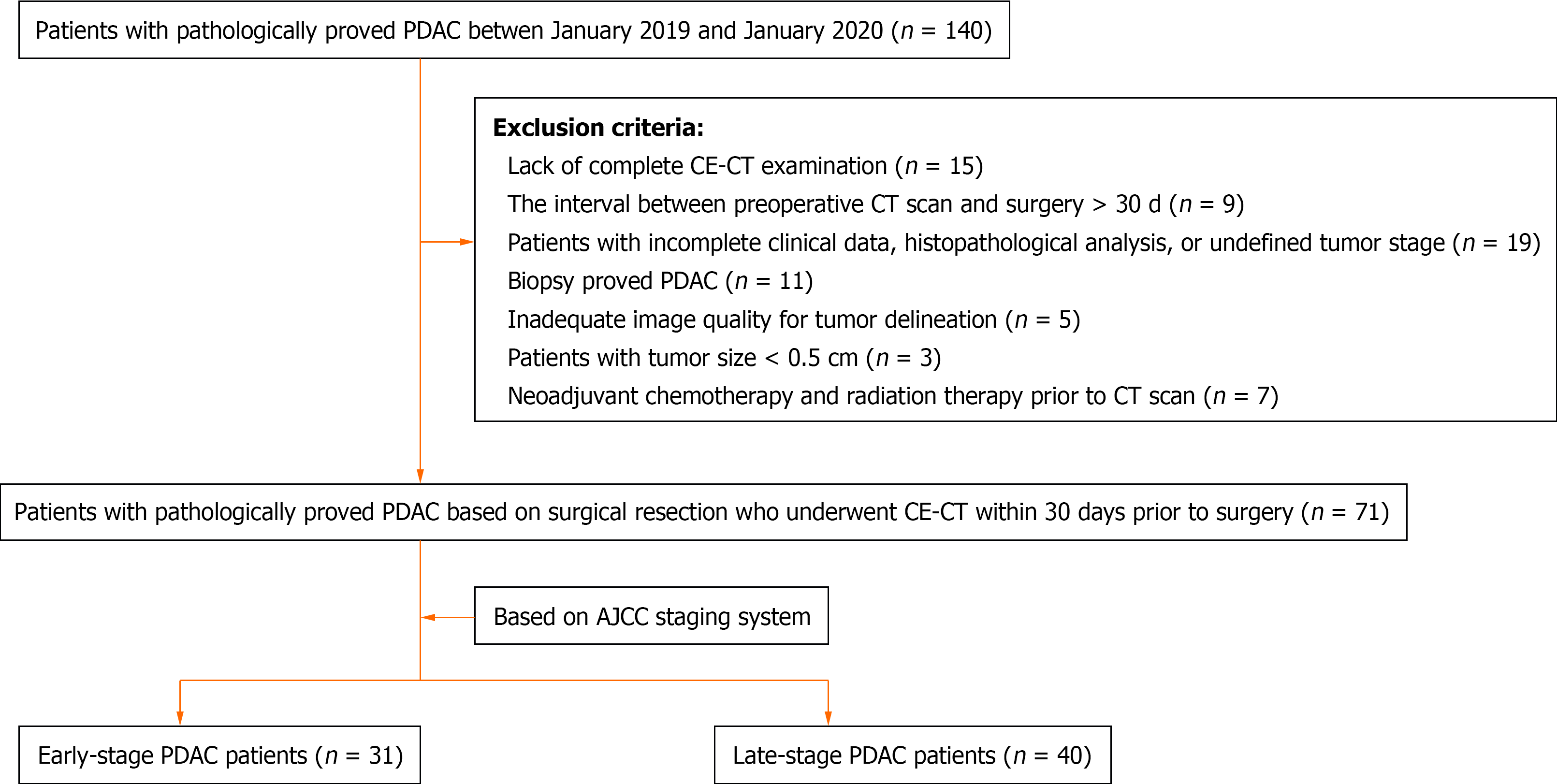
This study was approved by the ethics committee of Affiliated Hospital of Nanjing University of Chinese Medicine with waiver of informed consent due to its retrospective nature. The study was reviewed and approved by the ethics committee of Affiliated Hospital of Nanjing University of Chinese Medicine (Approval No. 2017NL-137-05). A total of 140 patients with pathologically proved PDAC from January 2019 to January 2020 was included in the study. Exclusion criteria were lack of complete contrast-enhanced CT examination (unenhanced, late arterial, and portal phases) (n = 15), the interval between preoperative CT scan and surgery > 30 d (n = 9), patients with incomplete clinical data, histopathological analysis, or undefined tumor stage (n = 19), biopsy proved PDAC (n = 11), inadequate image quality for tumor delineation (n = 5), patients with tumor size < 0.5 cm (n = 3), and neoadjuvant chemotherapy and radiation therapy prior to CT scan (n = 7). Finally, 71 patients with pathologically proved PDAC (39 males and 32 females with a mean age of 61.55 ± 8.53) were included in the study. Tumor staging was performed in accordance with the most current AJCC staging system (the 8th edition)[11]. A total of 71 patients were classified into stage IA (n = 4), stage IB (n = 8), stage IIA (n = 3), stage IIB (n = 16), stage III (n = 16) and stage IV (n = 24). Finally, patients with PDAC were defined as early-stage (I-II) and late stage (III-IV) (Figure 1).
All patients underwent triple-phase CT scan including unenhanced, late arterial, and portal venous phases. CT scanning was completed using one of the following scanners: (1) GE Optima 670 (GE Healthcare, Tokyo, Japan); (2) GE Lightspeed 64 VCT (GE Healthcare); (3) SIMENS SOMATOM Definition; and (4) Philips Brilliance 64 (Philips Healthcare, DA Best, the Netherlands). The following scan parameters were used: Tube voltage, 120Kvp; tube current, 200-400 mAs; helical pitch, 0.984-1.375; and 1.0 mm reconstruction slice thickness with an interval of 1.0 mm. An administration of 100-120 mL nonionic contrast media (Omnipaque 350, Bayer Pharmaceuticals) at a rate of 3.0-4.0 mL/s was performed after the unenhanced CT scan. The late arterial and portal venous phases were acquired at 35 s and 70 s, respectively.
The CT images were retrospectively reviewed by two abdominal radiologists in consensus. Any disparity was resolved by referring to a senior radiologist with 15 years’ experience of reading abdominal CT images. The following CT imaging features were evaluated in consensus: Tumor location, tumor margin (well-defined or ill-defined margin), presence of calcification, presence of cystic degeneration, presence of pancreatic duct dilatation, presence of vascular invasion, presence of lymph node metastasis, and presence of distal metastasis. Well-defined margin represents a margin that is smooth and visible, while spiculation or infiltration on one quarter of tumor perimeter indicates an ill-defined margin[17]. Calcifications were identified on unenhanced CT images. Cystic degeneration within the tumor was defined as low attenuation areas with CT attenuation values < 20 Hounsfield units or lack of enhancement on contrast-enhanced CT[18]. A maximal diameter ≥ 3 mm indicates pancreatic duct dilatation. Vascular invasion was defined in accordance with National Comprehensive Cancer Network guideline (version 2.2019-April 9, 2019)[19].
Dual-phase pancreatic CT protocol including late arterial and portal venous phases were recommended for the optimal evaluation of primary pancreatic tumors. In the present study, ITK-SNAP software (version 3.6.0) was used for tumor segmentation on the late arterial and portal venous phase images and regions of interest (ROIs) were manually outlined by the side of the complete tumor margin on all contiguous slices by an experienced radiologist, who was blinded to the histopathological and clinical findings of these patients and used ITK-SNAP for other studies consisting of 221 patients with confirmed pancreatic diseases[20,21].
Prior to feature extraction, gray-level normalization (a unified voxel size of 1.0 mm × 1.0 mm × 1.0 mm) was carried out in order to exclude disturbances caused by scanner variabilities and parameters before radiomics analysis[21]. A total of 792 radiomics features (396 from late arterial phase and 396 from portal venous phase) were extracted from the ROIs for each patient using Analysis Kit software (version V3.0.0.R, GE Healthcare). Mann-Whitney U test, univariate logistic regression analysis, and minimum redundancy maximum relevance (MRMR) method were used to identify the most pivotal and predictive radiomics features from all features in differential diagnosis between early-stage and late-stage PDAC.
The selected features from the late arterial and portal venous phases were used to construct the radiomics model using random forest (RF) method, which grows multiple decision trees that are merged together for a more accurate prediction. The prediction ability of the radiomics model was assessed and recorded.
The robustness and reproducibility of the radiomics model was validated using 10-times leave group out cross-validation (LGOCV) method. Patients were randomly divided into training and validation sets at a ratio of 7:3 for 10 times. For each time, a radiomics model was generated in the training set and the validation set was used to evaluate the predictive ability of the radiomics model. The robustness and reproducibility of the radiomics model was shown by the average performance of the 10 newly built models.
Statistical analysis was performed using SPSS v.24 (IBM Corp., Chicago, LA, United States) and R software v.3.6.1. P < 0.05 was considered to be statistically significant.
A total of 71 PDAC patients with a mean age of 61.55 ± 8.53 years were classified into early-stage (n = 31) and late-stage (n = 40) based on AJCC TNM staging criteria. The demographic characteristics were listed in Table 1. No statistically significant difference was found with respect to patients’ age (61.45 ± 8.93 years vs 61.63 ± 8.31 years, P = 0.933), sex, or demographic characteristics.
| Characteristic | Clinical stage classification | P value | |
| Early-stage PDAC (n = 31) | Late-stage PDAC (n = 40) | ||
| Age (yr) | 61.45 ± 8.93 | 61.63 ± 8.31 | 0.933 |
| Sex | 0.153 | ||
| Male | 20 (64.5%) | 19 (47.5%) | |
| Female | 11 (35.5%) | 21 (52.5%) | |
| Abdominal pain | 14 (45.2%) | 20 (50%) | 0.686 |
| Abdominal bloating | 8 (25.8%) | 11 (27.5) | 0.873 |
| Abdominal discomfort | 16 (51.6%) | 21 (52.5%) | 0.941 |
| Body weight loss | 11 (35.5%) | 14 (35.0%) | 0.966 |
| Jaundice | 7 (22.6%) | 9 (22.5) | 0.994 |
| AJCC staging | - | ||
| IA | 4 (12.9%) | ||
| IB | 8 (25.8%) | ||
| IIA | 3 (9.7%) | ||
| IIB | 16 (51.6%) | ||
| III | 0 | 16 (40%) | |
| IV | 0 | 24 (60%) | |
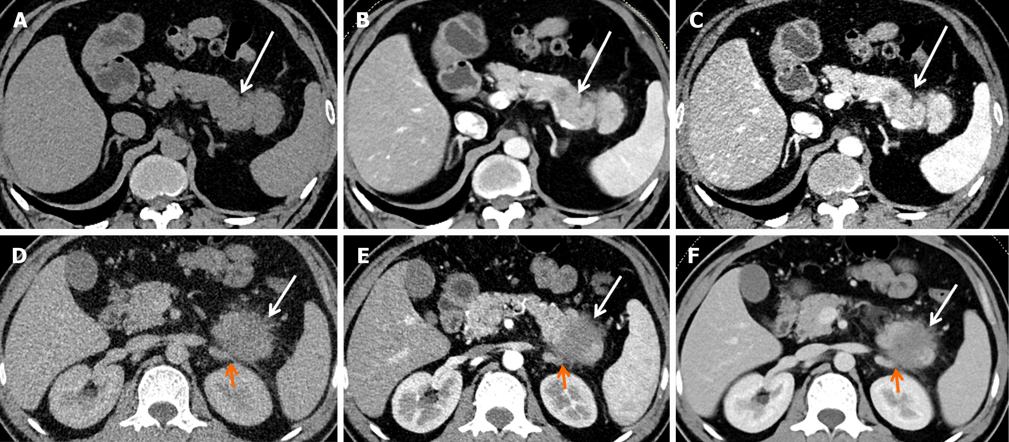
CT findings of patients with early-stage and late-stage PDAC were listed in Table 2. No statistically significant difference was found with respect to tumor location, tumor margin, calcification, cystic degeneration, pancreatic duct dilatation, vascular invasion, or lymph node metastasis. Late-stage PDAC has a larger tumor size as compared to early-stage PDAC (4.03 ± 1.25 cm vs 3.05 ± 0.54 cm, P < 0.001). Late-stage PDAC has a higher frequency of distal metastasis as compared to early-stage PDAC [24 (60%) vs 0 (0%), P < 0.001]. Two representative cases of early-stage and late-stage PDAC were shown in Figure 2. Case 1 represents a patient with stage IB PDAC, which located at the tail of the pancreas (Figure 2A-C). Case 2 represents a patient with stage IV PDAC, which located at the tail of the pancreas (Figure 2D-F).
| Characteristic | Clinical stage classification | P value | |
| Early-stage PDAC (n = 31) | Late-stage PDAC (n = 40) | ||
| Tumor size (cm) | 4.03 ± 1.25 | 3.05 ± 0.54 | < 0.001 |
| Tumor location | 0.869 | ||
| Head & neck | 18 (58.1%) | 24 (60.0%) | |
| Body & tail | 13 (41.9%) | 16 (40.0%) | |
| Tumor margin | 0.722 | ||
| Well-defined | 4 (12.9%) | 3 (7.5%) | |
| Ill-defined | 27 (87.1%) | 37 (92.5%) | |
| Calcification | 1.0 | ||
| Y | 1 (3.2%) | 1 (2.5%) | |
| N | 30 (96.8%) | 39 (97.5%) | |
| Cystic degeneration | 0.757 | ||
| Y | 3 (9.7%) | 6 (15.0%) | |
| N | 28 (90.3%) | 34 (85.0%) | |
| MPD | 0.861 | ||
| Y | 20 (64.5) | 25 (62.5%) | |
| N | 11 (35.5) | 15 (37.5%) | |
| Vascular invasion | 0.302 | ||
| Y | 14 (45.2%) | 23 (57.5%) | |
| N | 17 (54.8%) | 17 (42.5%) | |
| Lymph node metastasis | 0.480 | ||
| Y | 16 (51.6%) | 24 (60.0%) | |
| N | 15 (48.4%) | 16 (40.0%) | |
| Distal metastasis | < 0.001 | ||
| Y | 0 (0%) | 24 (60%) | |
| N | 31 (100%) | 16 (40%) | |
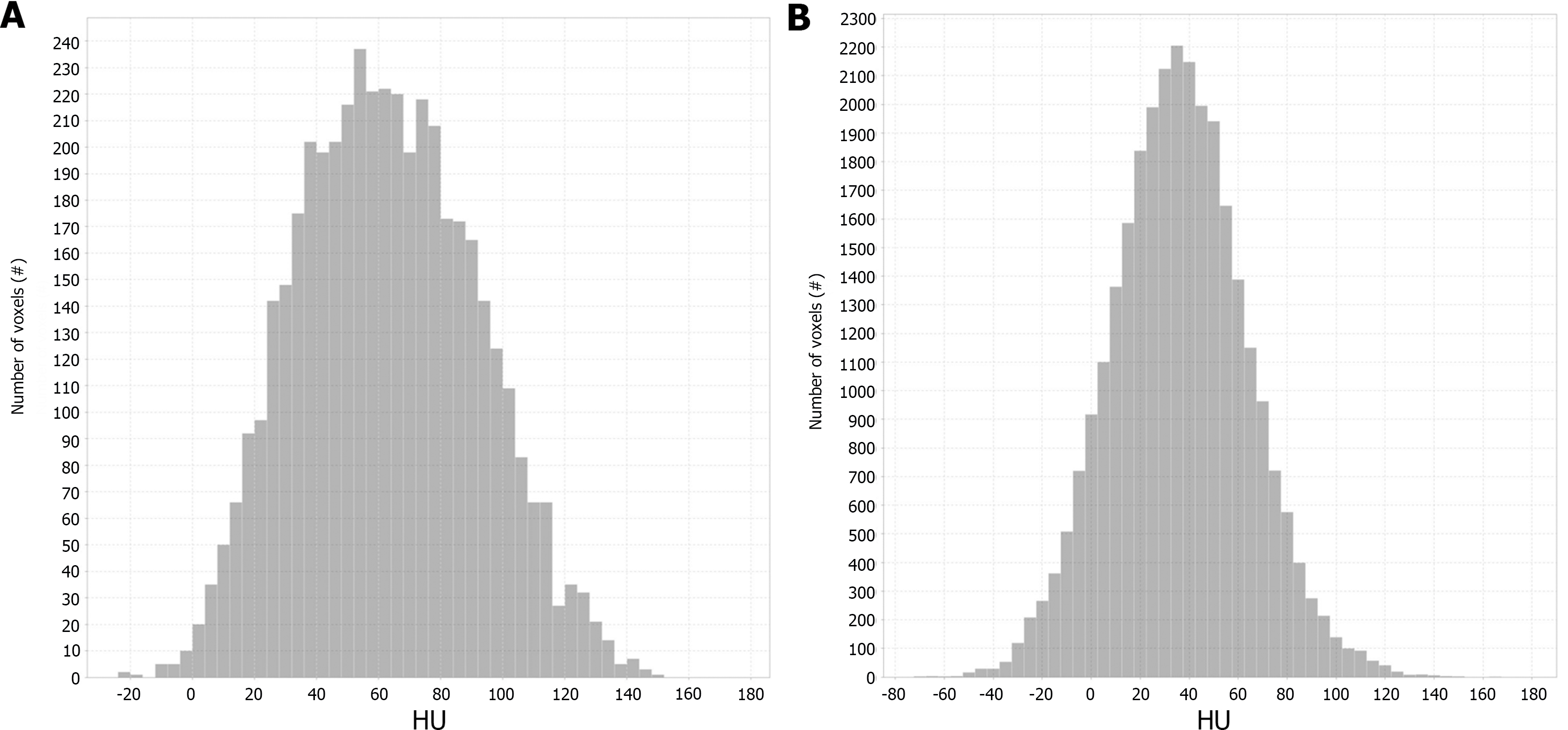
The extracted radiomics features from each phase CT imaging included histogram features (n = 42), morphological features (n = 9), grey level co-occurrence matrix (GLCM) features (n = 144), grey level size zone matrix features (n = 11), grey level run-length matrix features (n = 180), and Haralick features (n = 10).
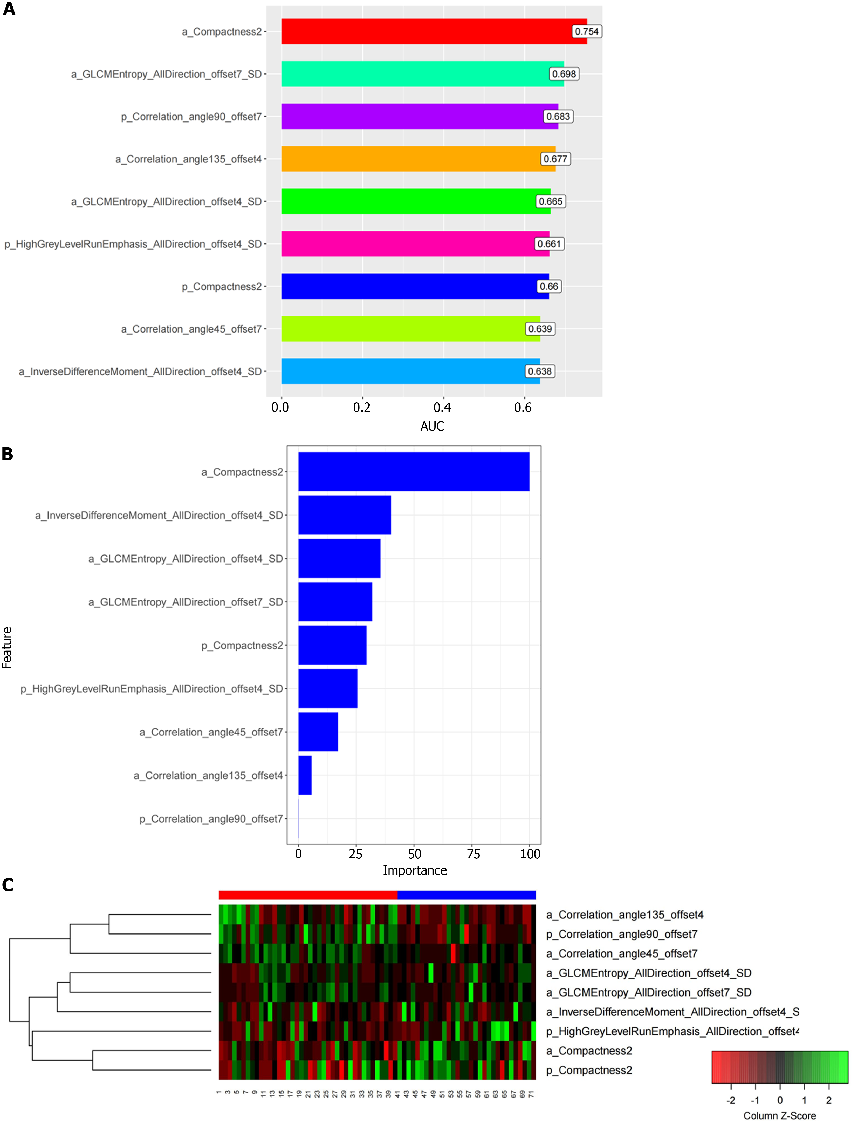
Nine most important and predictive radiomics features were identified after feature selection using Mann-Whitney U test, univariate logistic regression analysis, and MRMR method. Among them, 6 features were extracted from the late arterial phase (named a _ feature) and the others from the portal venous phase (named p _ feature). The selected features were a_Correlation_angle135_offset4, p_Correlation_angle90_offset7, a_Correlation_angle45_offset7, a_GLCM
| Features | AUC | SEN | SPE | ACC |
| a_Compactness2 | 0.754 | 67.5% | 77.42% | 71.83% |
| a_GLCMEntropy_AllDirection_offset7_SD | 0.698 | 82.5% | 58.06% | 71.83% |
| p_Correlation_angle90_offset7 | 0.683 | 60.0% | 70.97% | 64.79% |
| p_HighGreyLevelRunEmphasis_AllDirection_offset4_SD | 0.661 | 77.5% | 51.61% | 66.20% |
| a_GLCMEntropy_AllDirection_offset4_SD | 0.665 | 87.5% | 54.84% | 73.24% |
| a_Correlation_angle45_offset7 | 0.639 | 50.0% | 77.42% | 61.97% |
| p_Compactness2 | 0.66 | 80.0% | 54.84% | 69.01% |
| a_InverseDifferenceMoment_AllDirection_offset4_SD | 0.638 | 75.0% | 64.52% | 70.42% |
| a_Correlation_angle135_offset4 | 0.677 | 75.0% | 58.06% | 67.61% |
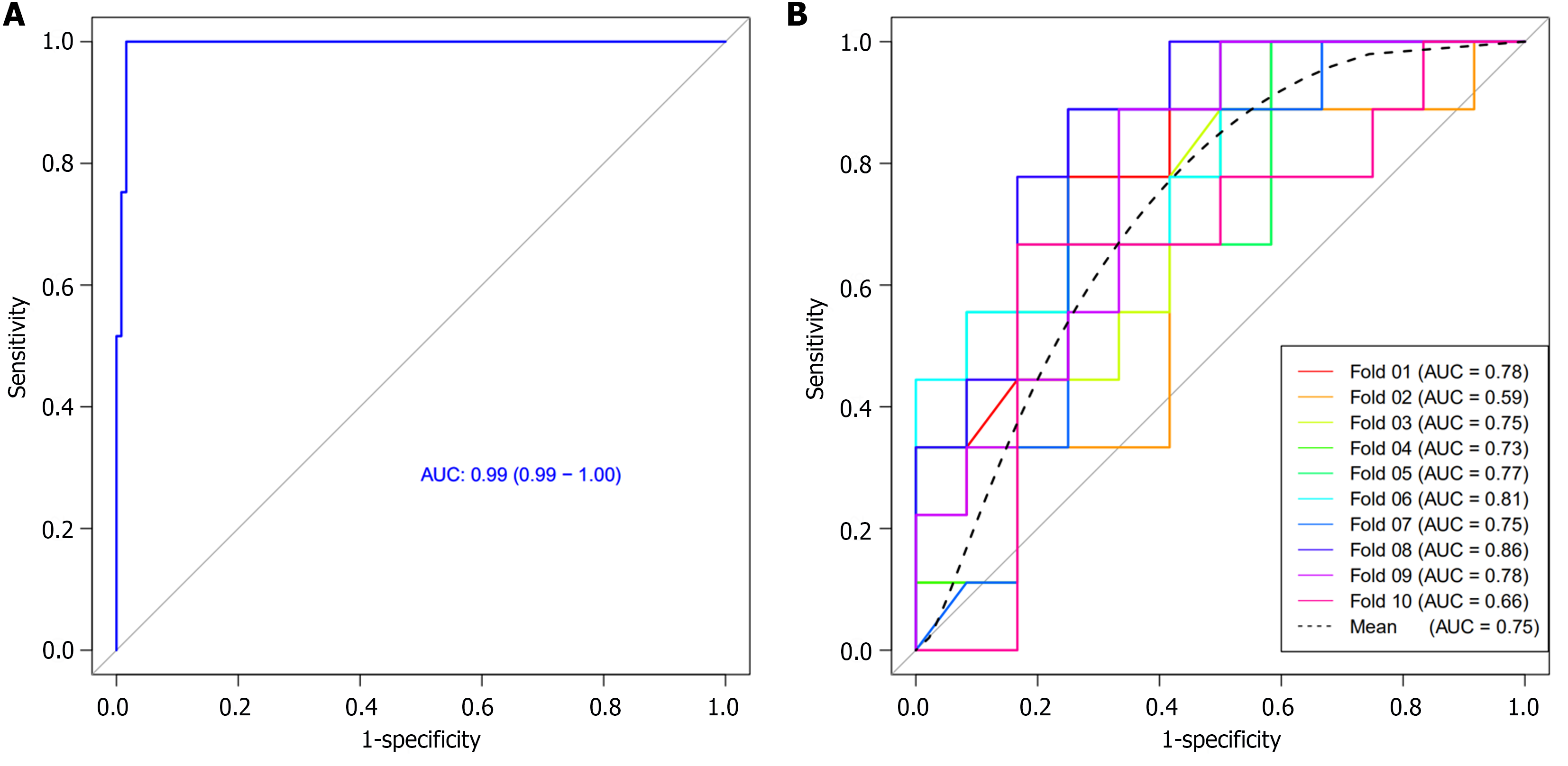
We subsequently adopted RF method to construct a radiomics model to differentiate between early-stage and late-stage PDAC, which had an AUC of 0.99 (Figure 5A). The RF classifier showed a high discriminative ability with 97.7% accuracy, 97.6% sensitivity, 97.8% specificity, 98.4% positive predictive value, and 96.8% negative predictive value. We finally used 10-times LGOCV method to validate the robustness and reproducibility of the radiomics model, with a mean AUC of 0.75 by the average performance of the 10 newly built models (Figure 5B), indicating good predictive ability of the radiomics model.
In the present study, we developed and validated a radiomics model in differentiating early-stage PDAC from late-stage PDAC. The radiomics model incorporating the most important and predictive features selected using Mann-Whitney U test, univariate logistic regression analysis, and MRMR method, displayed a high discriminative ability. The results demonstrated that radiomics analysis may facilitate the preoperative risk stratification, clinical decision making, and maximize patient survival in patients with PDAC with short life expectancy.
The accurate staging of PDAC and assessment of surgical resectability is crucial in the management of this dismal disease[22]. Xu et al[16] classified PDAC into early (stage I/II)- and late-stage (stage III/IV) and evaluated the diagnostic ability of a circRNA-based biomarker panel in distinguishing between them. In our study, patients with PDAC were also grouped as early-stage (I-II) and late stage (III-IV). No statistically significant difference was found with respect to patients’ age, sex, or demographic characteristics between early-stage and late-stage PDAC. This implied that early-stage and late-stage PDAC could not be differentiated by the demographic characteristics. The importance of tumor size was further underlined in the latest version of AJCC, especially for further grouping of T1-stage PDAC[22]. After CT imaging interpretation, late-stage PDAC has a larger tumor size as compared to early-stage PDAC (4.03 ± 1.25 cm vs 3.05 ± 0.54 cm, P < 0.001). Additionally, late-stage PDAC has a higher frequency of distal metastasis as compared to early-stage PDAC [24 (60%) vs 0 (0%), P < 0.001] since 24 patients with stage IV PDAC were included in late-stage PDAC group. Similarly, the sensitivity is low since stage III PDAC does not occur dismal metastasis.
Recently, some researchers have investigated the potential value of CT texture analysis in predicting the overall survival of PDAC patients and evaluating the resectability of PDAC[23]. Our latest research study also investigated the potential value of CT texture analysis for prediction of histopathological grade of PDAC[24]. Fifty-six PDAC patients were divided into low-grade and high-grade; the CT texture analysis based on support vector machine achieved 78% sensitivity, 95% specificity, and 86% accuracy. In the present study, we developed and validated a radiomics model in differentiating early-stage PDAC from late-stage PDAC. A total of 792 radiomics features (396 from the late arterial phase and 396 from the portal venous phase) were extracted from the ROI for each case. Nine most important and predictive radiomics features were identified after feature selection using Mann-Whitney U test, univariate logistic regression analysis, and MRMR method. Among which, 6 features were extracted from the late arterial phase and the others from the portal venous phase. RF method was used to construct a radiomics model, which shows a high predictive ability with an AUC of 0.99; subsequently, 10-times LGOCV method was used to validate the robustness and reproducibility of the radiomics model, with a mean AUC of 0.75 by the average performance of the 10 newly built models, indicating good predictive ability of the radiomics model.
There were several limitations in this study. First, the weak point of this paper is that the methods are very difficult for clinicians, even radiologists, to understand[21]. Therefore, specialized knowledge is required to understand radiomics or radscores. However, current radiomics work was dominated by analysis of semantic, radiologist-defined features and carried qualitative real-world meaning. Radiomics is similar to a black box in that only a fraction of the features can be interpreted at present. Second, the enrolled number of patients was relatively small due to the strict inclusion criteria. Multicenter validated prospective studies would be necessary to validate these promising outcomes. Third, the retrospective nature of this study inherently introduces a selection bias. However, the fact is that most research regarding texture analysis or machine learning is retrospective in nature[24]. Forth, this study only investigates the potential value of radiomics in PDAC staging differential diagnosis; the role of other imaging techniques including MRI and postoperative radiotherapy/MR has not been discussed. This is mainly due to the fact that CT-based radiomics to predict the histological grade of PDAC is more conducive to generalization since it only uses CT which is fast, low cost, and widely available for processing without additional expenses. Additionally, the National Comprehensive Cancer Network guideline recommends serial CT with contrast for routine follow-up to determine therapeutic benefit.
In conclusion, we developed and validated a radiomics model in differentiating early-stage PDAC from late-stage PDAC. The radiomics model incorporating the most important and predictive features had a high discriminative ability.
Pancreatic ductal adenocarcinoma (PDAC) remains the deadliest of the common cancers, with little change in patient survival in the past several decades. One of the biggest challenges of the management of PDAC that physicians often encounter is that the early detection in high-risk individuals and the early diagnosis of patients with suspected symptoms. Precise staging of PDAC is vital not only in making treatment decisions, but also in evaluating prognosis.
Radiomics, the generation of minable high throughput data through conversion of digital computed tomography (CT) or magnetic resonance imaging images, allows obtaining additional insight into pancreatic tissue heterogeneity. CT-based radiomics diagnostic approach could serve as a promising non-invasive method in differential diagnosis between early- and late-stage PDAC.
This study aimed to develop a radiomics-based diagnostic approach with a robust noninvasive diagnostic potential for identifying patients with early-stage PDAC.
A total of 71 patients with pathologically proved PDAC based on surgical resection who underwent contrast-enhanced-CT within 30 d prior to surgery were included in the study. Radiomics features were extracted from the region of interest (ROI) for each patient using Analysis Kit software. The most important and predictive radiomics features were selected using Mann-Whitney U test, univariate logistic regression analysis, and minimum redundancy maximum relevance (MRMR) method. Random forest (RF) method was used to construct the radiomics model, and 10-times leave group out cross-validation (LGOCV) method was used to validate the robustness and reproducibility of the model.
A total of 792 radiomics features (396 from late arterial phase and 396 from portal venous phase) were extracted from the ROI for each patient. Nine most important and predictive features were selected using Mann-Whitney U test, univariate logistic regression analysis, and MRMR method. RF method was used to construct the radiomics model with the nine most predictive radiomics features, which showed a high discriminative ability with 97.7% accuracy, 97.6% sensitivity, 97.8% specificity, 98.4% positive predictive value, and 96.8% negative predictive value. The radiomics model was proved to be robust and reproducible using 10-times LGOCV method with an average area under the curve of 0.75 by the average performance of the 10 newly built models.
This study demonstrated that CT-based radiomics diagnostic approach could be used to differentiate between early- and late-stage PDAC.
This study developed a radiomics-based diagnostic approach with a robust noninvasive diagnostic potential for identifying patients with early-stage PDAC. Large-scale prospective cohort studies, preferably multi-center, to validate the potential value of the radiomics diagnostic approach in differentiating early from late stage PDAC are in order.
We thank all authors for their continuous and excellent support with patient data collection, imaging analysis, statistical analysis and valuable suggestions for the article.
Provenance and peer review: Invited article; Externally peer reviewed.
Peer-review model: Single blind
Specialty type: Oncology
Country/Territory of origin: China
Peer-review report’s scientific quality classification
Grade A (Excellent): 0
Grade B (Very good): 0
Grade C (Good): C, C
Grade D (Fair): 0
Grade E (Poor): 0
P-Reviewer: Zeng C, United States S-Editor: Wang JJ L-Editor: A P-Editor: Cai YX
| 1. | Siegel RL, Miller KD, Wagle NS, Jemal A. Cancer statistics, 2023. CA Cancer J Clin. 2023;73:17-48. [RCA] [PubMed] [DOI] [Full Text] [Cited by in Crossref: 116] [Cited by in RCA: 9838] [Article Influence: 4919.0] [Reference Citation Analysis (2)] |
| 2. | Miller FH, Lopes Vendrami C, Hammond NA, Mittal PK, Nikolaidis P, Jawahar A. Pancreatic Cancer and Its Mimics. Radiographics. 2023;43:e230054. [RCA] [PubMed] [DOI] [Full Text] [Cited by in RCA: 16] [Reference Citation Analysis (0)] |
| 3. | Guler GD, Ning Y, Ku CJ, Phillips T, McCarthy E, Ellison CK, Bergamaschi A, Collin F, Lloyd P, Scott A, Antoine M, Wang W, Chau K, Ashworth A, Quake SR, Levy S. Detection of early stage pancreatic cancer using 5-hydroxymethylcytosine signatures in circulating cell free DNA. Nat Commun. 2020;11:5270. [RCA] [PubMed] [DOI] [Full Text] [Full Text (PDF)] [Cited by in Crossref: 89] [Cited by in RCA: 114] [Article Influence: 22.8] [Reference Citation Analysis (0)] |
| 4. | Nagai M, Nakamura K, Terai T, Kohara Y, Yasuda S, Matsuo Y, Doi S, Sakata T, Sho M. Significance of multiple tumor markers measurements in conversion surgery for unresectable locally advanced pancreatic cancer. Pancreatology. 2023;23:721-728. [RCA] [PubMed] [DOI] [Full Text] [Reference Citation Analysis (0)] |
| 5. | Kim SS, Lee S, Lee HS, Bang S, Han K, Park MS. Retrospective Evaluation of Treatment Response in Patients with Nonmetastatic Pancreatic Cancer Using CT and CA 19-9. Radiology. 2022;303:548-556. [RCA] [PubMed] [DOI] [Full Text] [Cited by in Crossref: 9] [Cited by in RCA: 9] [Article Influence: 3.0] [Reference Citation Analysis (0)] |
| 6. | Coppola A, La Vaccara V, Fiore M, Farolfi T, Ramella S, Angeletti S, Coppola R, Caputo D. CA19.9 Serum Level Predicts Lymph-Nodes Status in Resectable Pancreatic Ductal Adenocarcinoma: A Retrospective Single-Center Analysis. Front Oncol. 2021;11:690580. [RCA] [PubMed] [DOI] [Full Text] [Full Text (PDF)] [Cited by in Crossref: 20] [Cited by in RCA: 16] [Article Influence: 4.0] [Reference Citation Analysis (0)] |
| 7. | Huang B, Huang H, Zhang S, Zhang D, Shi Q, Liu J, Guo J. Artificial intelligence in pancreatic cancer. Theranostics. 2022;12:6931-6954. [RCA] [PubMed] [DOI] [Full Text] [Full Text (PDF)] [Cited by in Crossref: 46] [Cited by in RCA: 52] [Article Influence: 17.3] [Reference Citation Analysis (0)] |
| 8. | Zhang L, Sanagapalli S, Stoita A. Challenges in diagnosis of pancreatic cancer. World J Gastroenterol. 2018;24:2047-2060. [RCA] [PubMed] [DOI] [Full Text] [Full Text (PDF)] [Cited by in CrossRef: 272] [Cited by in RCA: 369] [Article Influence: 52.7] [Reference Citation Analysis (12)] |
| 9. | Cheng H, Luo G, Jin K, Xiao Z, Qian Y, Gong Y, Yu X, Liu C. Predictive Values of Preoperative Markers for Resectable Pancreatic Body and Tail Cancer Determined by MDCT to Detect Occult Metastases. World J Surg. 2021;45:2185-2190. [RCA] [PubMed] [DOI] [Full Text] [Cited by in Crossref: 9] [Cited by in RCA: 4] [Article Influence: 1.0] [Reference Citation Analysis (0)] |
| 10. | Ahmed SA, Atta H, Hassan RA. The utility of Multi-Detector Computed Tomography criteria after neoadjuvant therapy in Borderline Resectable Pancreatic cancer: Prospective, bi-institutional study. Eur J Radiol. 2021;139:109685. [RCA] [PubMed] [DOI] [Full Text] [Cited by in Crossref: 7] [Cited by in RCA: 2] [Article Influence: 0.5] [Reference Citation Analysis (0)] |
| 11. | Kulkarni NM, Soloff EV, Tolat PP, Sangster GP, Fleming JB, Brook OR, Wang ZJ, Hecht EM, Zins M, Bhosale PR, Arif-Tiwari H, Mannelli L, Kambadakone AR, Tamm EP. White paper on pancreatic ductal adenocarcinoma from society of abdominal radiology's disease-focused panel for pancreatic ductal adenocarcinoma: Part I, AJCC staging system, NCCN guidelines, and borderline resectable disease. Abdom Radiol (NY). 2020;45:716-728. [RCA] [PubMed] [DOI] [Full Text] [Cited by in Crossref: 42] [Cited by in RCA: 31] [Article Influence: 6.2] [Reference Citation Analysis (0)] |
| 12. | Kulkarni NM, Mannelli L, Zins M, Bhosale PR, Arif-Tiwari H, Brook OR, Hecht EM, Kastrinos F, Wang ZJ, Soloff EV, Tolat PP, Sangster G, Fleming J, Tamm EP, Kambadakone AR. White paper on pancreatic ductal adenocarcinoma from society of abdominal radiology's disease-focused panel for pancreatic ductal adenocarcinoma: Part II, update on imaging techniques and screening of pancreatic cancer in high-risk individuals. Abdom Radiol (NY). 2020;45:729-742. [RCA] [PubMed] [DOI] [Full Text] [Cited by in Crossref: 24] [Cited by in RCA: 24] [Article Influence: 4.8] [Reference Citation Analysis (0)] |
| 13. | Berbís MÁ, Godino FP, Rodríguez-Comas J, Nava E, García-Figueiras R, Baleato-González S, Luna A. Radiomics in CT and MR imaging of the liver and pancreas: tools with potential for clinical application. Abdom Radiol (NY). 2024;49:322-340. [RCA] [PubMed] [DOI] [Full Text] [Cited by in RCA: 9] [Reference Citation Analysis (0)] |
| 14. | Xiao G, Rong WC, Hu YC, Shi ZQ, Yang Y, Ren JL, Cui GB. MRI Radiomics Analysis for Predicting the Pathologic Classification and TNM Staging of Thymic Epithelial Tumors: A Pilot Study. AJR Am J Roentgenol. 2020;214:328-340. [RCA] [PubMed] [DOI] [Full Text] [Cited by in Crossref: 15] [Cited by in RCA: 33] [Article Influence: 6.6] [Reference Citation Analysis (0)] |
| 15. | Tabari A, Chan SM, Omar OMF, Iqbal SI, Gee MS, Daye D. Role of Machine Learning in Precision Oncology: Applications in Gastrointestinal Cancers. Cancers (Basel). 2022;15. [RCA] [PubMed] [DOI] [Full Text] [Full Text (PDF)] [Cited by in Crossref: 2] [Cited by in RCA: 20] [Article Influence: 6.7] [Reference Citation Analysis (0)] |
| 16. | Xu C, Jun E, Okugawa Y, Toiyama Y, Borazanci E, Bolton J, Taketomi A, Kim SC, Shang D, Von Hoff D, Zhang G, Goel A. A Circulating Panel of circRNA Biomarkers for the Noninvasive and Early Detection of Pancreatic Ductal Adenocarcinoma. Gastroenterology. 2024;166:178-190.e16. [RCA] [PubMed] [DOI] [Full Text] [Cited by in Crossref: 18] [Cited by in RCA: 36] [Article Influence: 36.0] [Reference Citation Analysis (0)] |
| 17. | Ren S, Qian L, Daniels MJ, Duan S, Chen R, Wang Z. Evaluation of contrast-enhanced computed tomography for the differential diagnosis of hypovascular pancreatic neuroendocrine tumors from chronic mass-forming pancreatitis. Eur J Radiol. 2020;133:109360. [RCA] [PubMed] [DOI] [Full Text] [Cited by in Crossref: 13] [Cited by in RCA: 7] [Article Influence: 1.4] [Reference Citation Analysis (0)] |
| 18. | Ren S, Chen X, Wang J, Zhao R, Song L, Li H, Wang Z. Differentiation of duodenal gastrointestinal stromal tumors from hypervascular pancreatic neuroendocrine tumors in the pancreatic head using contrast-enhanced computed tomography. Abdom Radiol (NY). 2019;44:867-876. [RCA] [PubMed] [DOI] [Full Text] [Cited by in Crossref: 8] [Cited by in RCA: 5] [Article Influence: 0.8] [Reference Citation Analysis (0)] |
| 19. | Tempero MA, Malafa MP, Chiorean EG, Czito B, Scaife C, Narang AK, Fountzilas C, Wolpin BM, Al-Hawary M, Asbun H, Behrman SW, Benson AB, Binder E, Cardin DB, Cha C, Chung V, Dillhoff M, Dotan E, Ferrone CR, Fisher G, Hardacre J, Hawkins WG, Ko AH, LoConte N, Lowy AM, Moravek C, Nakakura EK, O'Reilly EM, Obando J, Reddy S, Thayer S, Wolff RA, Burns JL, Zuccarino-Catania G. Pancreatic Adenocarcinoma, Version 1.2019. J Natl Compr Canc Netw. 2019;17:202-210. [RCA] [PubMed] [DOI] [Full Text] [Cited by in Crossref: 194] [Cited by in RCA: 271] [Article Influence: 54.2] [Reference Citation Analysis (2)] |
| 20. | Ren S, Zhang J, Chen J, Cui W, Zhao R, Qiu W, Duan S, Chen R, Chen X, Wang Z. Evaluation of Texture Analysis for the Differential Diagnosis of Mass-Forming Pancreatitis From Pancreatic Ductal Adenocarcinoma on Contrast-Enhanced CT Images. Front Oncol. 2019;9:1171. [RCA] [PubMed] [DOI] [Full Text] [Full Text (PDF)] [Cited by in Crossref: 36] [Cited by in RCA: 37] [Article Influence: 6.2] [Reference Citation Analysis (0)] |
| 21. | Ren S, Zhao R, Cui W, Qiu W, Guo K, Cao Y, Duan S, Wang Z, Chen R. Computed Tomography-Based Radiomics Signature for the Preoperative Differentiation of Pancreatic Adenosquamous Carcinoma From Pancreatic Ductal Adenocarcinoma. Front Oncol. 2020;10:1618. [RCA] [PubMed] [DOI] [Full Text] [Full Text (PDF)] [Cited by in Crossref: 30] [Cited by in RCA: 31] [Article Influence: 6.2] [Reference Citation Analysis (0)] |
| 22. | Ma C, Yang P, Li J, Bian Y, Wang L, Lu J. Pancreatic adenocarcinoma: variability in measurements of tumor size among computed tomography, magnetic resonance imaging, and pathologic specimens. Abdom Radiol (NY). 2020;45:782-788. [RCA] [PubMed] [DOI] [Full Text] [Cited by in Crossref: 9] [Cited by in RCA: 8] [Article Influence: 1.6] [Reference Citation Analysis (0)] |
| 23. | Cassinotto C, Chong J, Zogopoulos G, Reinhold C, Chiche L, Lafourcade JP, Cuggia A, Terrebonne E, Dohan A, Gallix B. Resectable pancreatic adenocarcinoma: Role of CT quantitative imaging biomarkers for predicting pathology and patient outcomes. Eur J Radiol. 2017;90:152-158. [RCA] [PubMed] [DOI] [Full Text] [Cited by in Crossref: 64] [Cited by in RCA: 83] [Article Influence: 10.4] [Reference Citation Analysis (0)] |
| 24. | Qiu W, Duan N, Chen X, Ren S, Zhang Y, Wang Z, Chen R. Pancreatic Ductal Adenocarcinoma: Machine Learning-Based Quantitative Computed Tomography Texture Analysis For Prediction Of Histopathological Grade. Cancer Manag Res. 2019;11:9253-9264. [RCA] [PubMed] [DOI] [Full Text] [Full Text (PDF)] [Cited by in Crossref: 32] [Cited by in RCA: 31] [Article Influence: 5.2] [Reference Citation Analysis (0)] |


