Copyright
©The Author(s) 2017.
World J Gastrointest Oncol. Sep 15, 2017; 9(9): 397-401
Published online Sep 15, 2017. doi: 10.4251/wjgo.v9.i9.397
Published online Sep 15, 2017. doi: 10.4251/wjgo.v9.i9.397
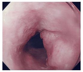
Figure 1 Esophagoscopic view of the tumor.
Esophagoscopy reveals a type 1 tumor in the cervical esophagus.
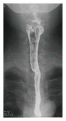
Figure 2 Upper gastrointestinal barium study image.
An upper gastrointestinal barium study reveals a 15-mm filling defect at the cervical esophagus.
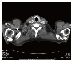
Figure 3 Preoperative computed tomography image.
Contrast-enhanced computed tomography indicates weakly enhanced swollen lymph nodes in the right cervical region.
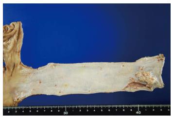
Figure 4 Surgical specimen of the esophagus.
Macroscopically, the type 1 tumor is located in the cervical esophagus, and it measures 30 mm × 15 mm.
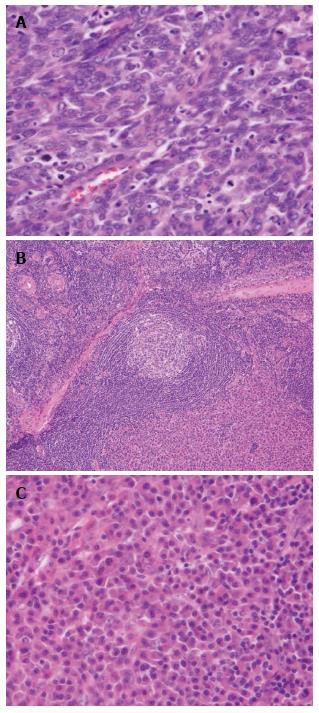
Figure 5 Histopathological results of the resected specimen.
A: The histopathological diagnosis of the resected esophagus is moderately differentiated squamous cell carcinoma (HE × 200); B: Histological examination of the right-sided neck lymph nodes reveals onionskin arrangement of small lymphocytes (HE × 20); C: Interfollicular diffuse proliferation of plasma cells (HE × 200).
- Citation: Yamabuki T, Ohara M, Kato M, Kimura N, Shirosaki T, Okamura K, Fujiwara A, Takahashi R, Komuro K, Iwashiro N, Hirano S. Cervical Castleman’s disease mimicking lymph node metastasis of esophageal carcinoma. World J Gastrointest Oncol 2017; 9(9): 397-401
- URL: https://www.wjgnet.com/1948-5204/full/v9/i9/397.htm
- DOI: https://dx.doi.org/10.4251/wjgo.v9.i9.397


