Copyright
©The Author(s) 2017.
World J Gastrointest Oncol. May 15, 2017; 9(5): 218-227
Published online May 15, 2017. doi: 10.4251/wjgo.v9.i5.218
Published online May 15, 2017. doi: 10.4251/wjgo.v9.i5.218
Figure 1 Total pelvic and lateral exenteration.
A: A Deaver retractor was placed caudally (White arrow: Pelvic tumour; Yellow arrow: Right obturator nerve; Blue and purple arrows: Right internal iliac vein and artery respectively; Green arrow: Transected right distal ureter at pelvic brim with infant feeding tube inserted for intraoperative urinary diversion); B: Post-exenteration view showing the right internal iliac vessels, obturator nerve and pelvic lymph nodes excised and the metal vacuum tube pointed at exposed pelvic bone (Yellow arrow: Sciatic nerve; Blue arrow: Right external iliac vessels; Green arrow: Transected right distal ureter); C: Cicatrising tumour specimen showing invasion into bladder and right pelvic sidewall (Blue arrow: Right internal iliac vessels).
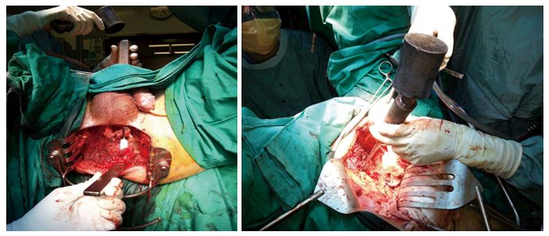
Figure 2 Demonstration of abdominal perineal approach for level S3 sacrectomy.
The left panel (perineal view) demonstrates placement of the osteotomes posterior to the sacral bone in order to protect perineal skin while the surgeon transects the sacrum at S3 level in the right panel (abdominal view).
- Citation: Chew MH, Yeh YT, Toh EL, Sumarli SA, Chew GK, Lee LS, Tan MH, Hennedige TP, Ng SY, Lee SK, Chong TT, Abdullah HR, Goh TLH, Rasheed MZ, Tan KC, Tang CL. Critical evaluation of contemporary management in a new Pelvic Exenteration Unit: The first 25 consecutive cases. World J Gastrointest Oncol 2017; 9(5): 218-227
- URL: https://www.wjgnet.com/1948-5204/full/v9/i5/218.htm
- DOI: https://dx.doi.org/10.4251/wjgo.v9.i5.218


