Copyright
©The Author(s) 2017.
World J Gastrointest Oncol. Nov 15, 2017; 9(11): 452-456
Published online Nov 15, 2017. doi: 10.4251/wjgo.v9.i11.452
Published online Nov 15, 2017. doi: 10.4251/wjgo.v9.i11.452
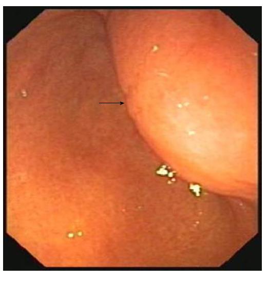
Figure 1 Upper gastrointestinal endoscopy revealing a subepithelial tumor with intact overlying mucosa (black arrow) on the posterior wall of the gastric antrum.
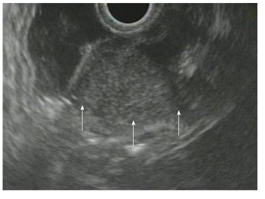
Figure 2 Endoscopic ultrasonography showing a homogenous, hypoechoic mass within the muscularis propria (white arrows).
Its echogenicity appears to be more hyperechoic than that of the muscle layer.
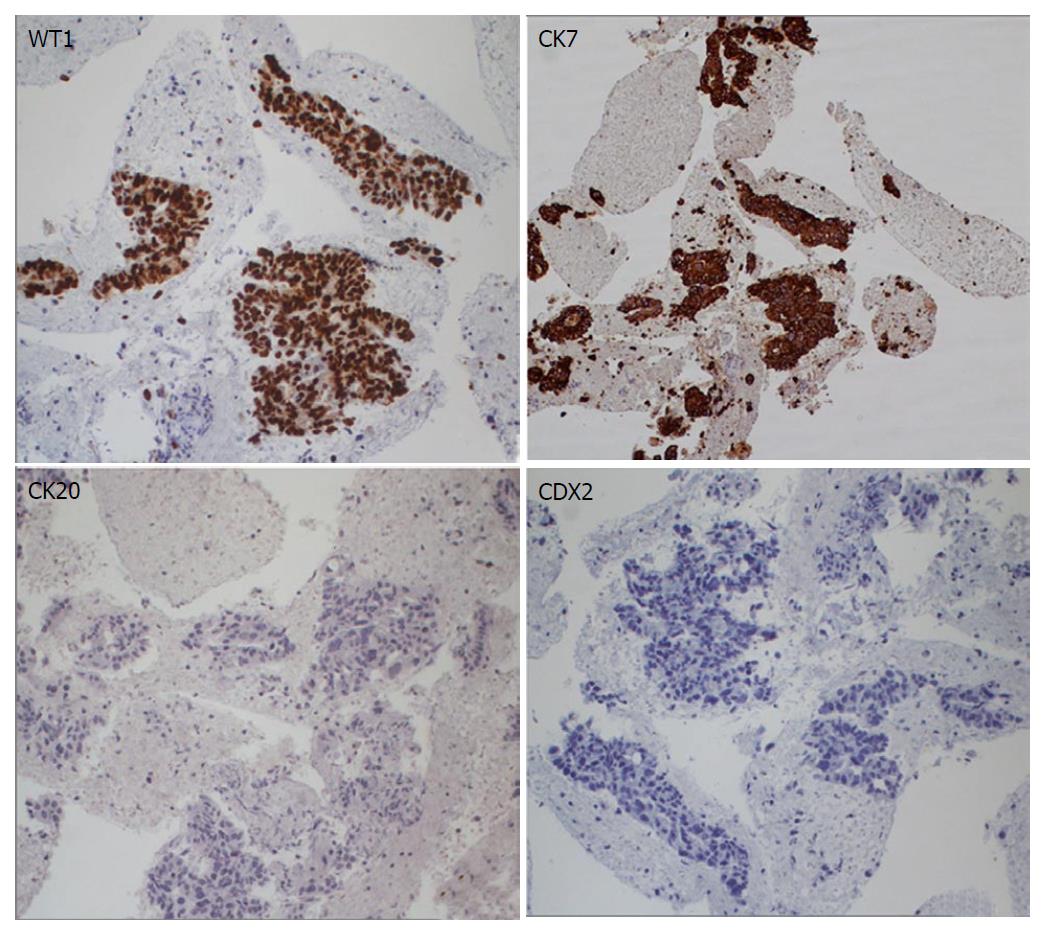
Figure 3 Histopathological images showing adenocarcinoma with immunohistochemistry positive for WT1 (× 20) and CK7 (× 16), and negative for CK20 (× 20) and CDX2 (× 20).
- Citation: Antonini F, Laterza L, Fuccio L, Marcellini M, Angelelli L, Calcina S, Rubini C, Macarri G. Gastric metastasis from ovarian adenocarcinoma presenting as a subepithelial tumor and diagnosed by endoscopic ultrasound-guided tissue acquisition. World J Gastrointest Oncol 2017; 9(11): 452-456
- URL: https://www.wjgnet.com/1948-5204/full/v9/i11/452.htm
- DOI: https://dx.doi.org/10.4251/wjgo.v9.i11.452


