Copyright
©The Author(s) 2017.
World J Gastrointest Oncol. Oct 15, 2017; 9(10): 431-435
Published online Oct 15, 2017. doi: 10.4251/wjgo.v9.i10.431
Published online Oct 15, 2017. doi: 10.4251/wjgo.v9.i10.431
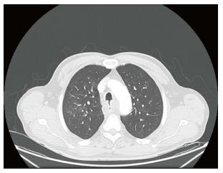
Figure 1 Contrasted chest computed tomography imaging showing tracheoesophageal fistula in a 60-year-old male patient.
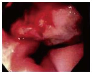
Figure 2 Esophagogastroduodenoscopy showing a partially obstructing mid-esophageal tumor and tracheoesophageal fistula in a 60-year-old male patient.
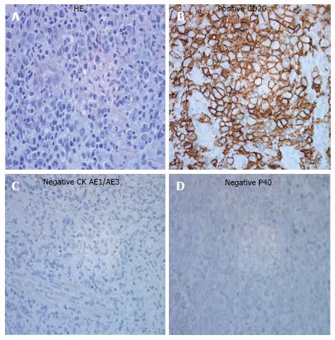
Figure 3 Histological features of primary diffuse large B-cell lymphoma in a 60-year-old male patient.
A: HE staining shows highly pleomorphic large cell proliferation on sections of neoplasm; B: Immunohistochemistry shows tumor cells with a strongly diffused positive expression for CD20; C: Cytokeratin (CK) AE1/AE3 was negative for the cells of tumor infiltrate; D: P40 was negative for squamous carcinoma.
- Citation: Teerakanok J, DeWitt JP, Juarez E, Thein KZ, Warraich I. Primary esophageal diffuse large B cell lymphoma presenting with tracheoesophageal fistula: A rare case and review. World J Gastrointest Oncol 2017; 9(10): 431-435
- URL: https://www.wjgnet.com/1948-5204/full/v9/i10/431.htm
- DOI: https://dx.doi.org/10.4251/wjgo.v9.i10.431


