Copyright
©The Author(s) 2017.
World J Gastrointest Oncol. Oct 15, 2017; 9(10): 416-422
Published online Oct 15, 2017. doi: 10.4251/wjgo.v9.i10.416
Published online Oct 15, 2017. doi: 10.4251/wjgo.v9.i10.416
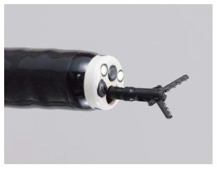
Figure 1 The distal tip of the Clutch Cutter (long type: Blade length of 5 mm).
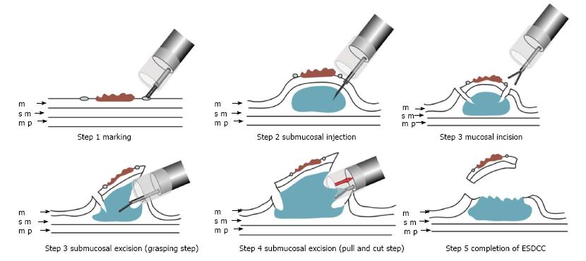
Figure 2 Schema showing endoscopic submucosal dissection using the Clutch Cutter technique.
m: Mucosa; sm: Submucosa; mp: Muscularis propria; ESDCC: Endoscopic submucosal dissection using the Clutch Cutter.
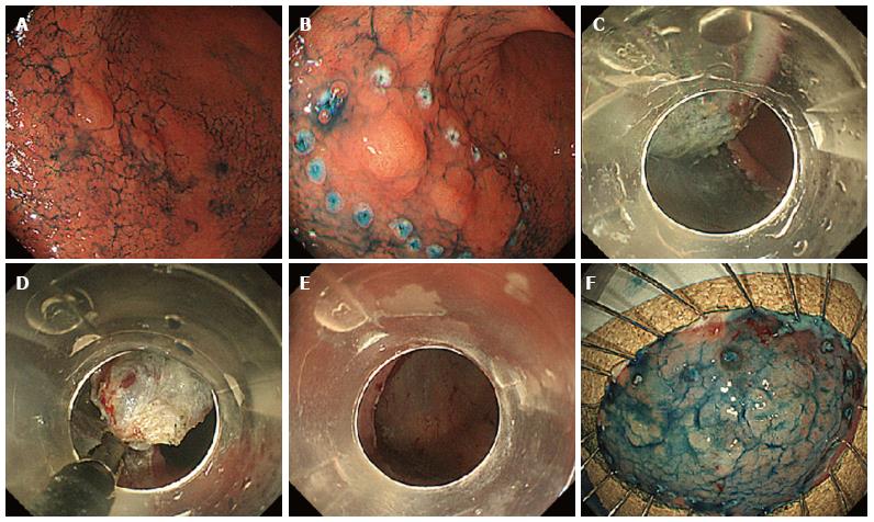
Figure 3 Endoscopic submucosal dissection using the Clutch Cutter in an 82-year-old Japanese male.
A: Indigo carmine was sprayed to demarcate the lesion; B: Markings outside the lesion; C and D: The submucosal tissue under the lesion was gradually grasped and dissected from the muscle layer; E: The lesion was completely cut from the muscle layer; F: Fixation of the specimen.
- Citation: Otsuka Y, Akahoshi K, Yasunaga K, Kubokawa M, Gibo J, Osada S, Tokumaru K, Miyamoto K, Sato T, Shiratsuchi Y, Oya M, Koga H, Ihara E, Nakamura K. Clinical outcomes of Clutch Cutter endoscopic submucosal dissection for older patients with early gastric cancer. World J Gastrointest Oncol 2017; 9(10): 416-422
- URL: https://www.wjgnet.com/1948-5204/full/v9/i10/416.htm
- DOI: https://dx.doi.org/10.4251/wjgo.v9.i10.416


