Copyright
©The Author(s) 2017.
World J Gastrointest Oncol. Jan 15, 2017; 9(1): 21-29
Published online Jan 15, 2017. doi: 10.4251/wjgo.v9.i1.21
Published online Jan 15, 2017. doi: 10.4251/wjgo.v9.i1.21
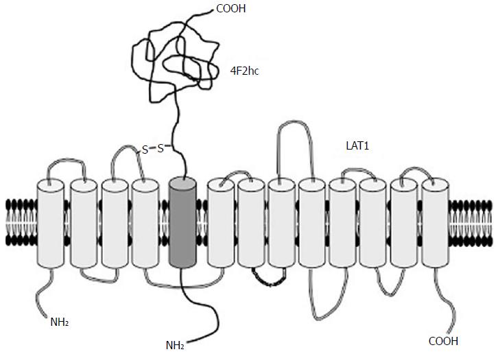
Figure 1 Structure of LAT1.
LAT1 is composed of 12 transmembrane helices that are predicted to form a cylindrical conformation penetrating the cellular membrane. LAT1 associates with 4F2hc for stable localization at the cellular membrane. LAT2 is similar in structure to LAT1, whereas LAT3 and LAT4 function independently of 4F2hc.
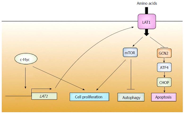
Figure 2 Schematic model of acquisition and monitoring of amino acids in cancer.
c-Myc promotes expression of LAT1, which supplies amino acids necessary for growth of cancers. The availability of amino acids is constantly monitored by factors such as mTOR and GCN2. Once amino acid deficiency is detected, cancers suppress their proliferation and, as occasion demands, induce apoptosis. mTOR: Mechanistic target of rapamycin; GCN2: General control non-derepressible 2; ATF4: Activating transcription factor 4; CHOP: C/EBP homologous protein.
- Citation: Hayashi K, Anzai N. Novel therapeutic approaches targeting L-type amino acid transporters for cancer treatment. World J Gastrointest Oncol 2017; 9(1): 21-29
- URL: https://www.wjgnet.com/1948-5204/full/v9/i1/21.htm
- DOI: https://dx.doi.org/10.4251/wjgo.v9.i1.21


