Copyright
©The Author(s) 2015.
World J Gastrointest Oncol. Mar 15, 2015; 7(3): 12-16
Published online Mar 15, 2015. doi: 10.4251/wjgo.v7.i3.12
Published online Mar 15, 2015. doi: 10.4251/wjgo.v7.i3.12
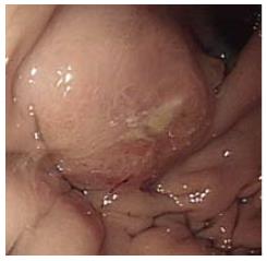
Figure 1 Esophagogastroduodenoscopy shows a Bormann type IV gastric mucosal lesion and loss of distension of the gastric wall.
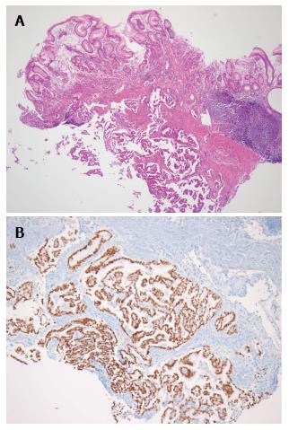
Figure 2 Hematoxylin-and-eosin (× 40) staining of the gastric lesion shows adenocarcinoma cells infiltrating the gastric submucosa (A) and thyroid transcription factor-1 positive staining in the cancerous gastric lesion (× 100) (B).
- Citation: Kim MJ, Hong JH, Park ES, Byun JH. Gastric metastasis from primary lung adenocarcinoma mimicking primary gastric cancer. World J Gastrointest Oncol 2015; 7(3): 12-16
- URL: https://www.wjgnet.com/1948-5204/full/v7/i3/12.htm
- DOI: https://dx.doi.org/10.4251/wjgo.v7.i3.12


