Copyright
©2014 Baishideng Publishing Group Inc.
World J Gastrointest Oncol. Sep 15, 2014; 6(9): 377-380
Published online Sep 15, 2014. doi: 10.4251/wjgo.v6.i9.377
Published online Sep 15, 2014. doi: 10.4251/wjgo.v6.i9.377
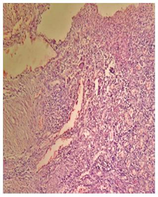
Figure 1 Histology showed extensive mucosal necrosis surrounded by lymphocytes and langhans giant cells.
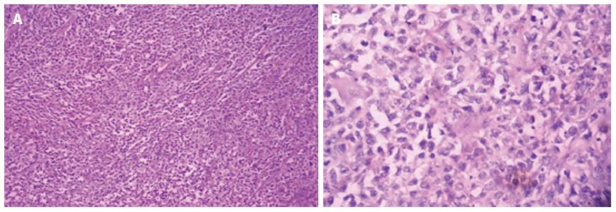
Figure 2 Histology showed moderately differentiated adenocarcinoma infiltrating the submucosa and serosa (A: HE, × 10) with mitotic figures and surrounded by neoplastic lymphocytes (B: HE, × 40).
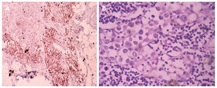
Figure 3 Mucosa associated lymphoid tissue lymphoma showing CD20 positivity (A: IHC, × 4) and Diffuse cytoplasmic positivity of cytokeratin in pericolonic tissue suggestive of adenocarcinoma (B: IHC, × 40).
Inconvenience regretted. Kindly make the necessary change.
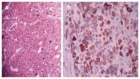
Figure 4 Mucosa associated lymphoid tissue lymphoma showing CD20 positivity(A: IHC, × 4) and diffuse cytoplasmic positivity of cytokeratin in pericolonic tissue suggestive of adenocarcinoma(B: IHC, × 40).
- Citation: Velu ARK, Srinivasamurthy BC, Nagarajan K, Sinduja I. Colonic adenocarcinoma, mucosa associated lymphoid tissue lymphoma and tuberculosis in a segment of colon: A case report. World J Gastrointest Oncol 2014; 6(9): 377-380
- URL: https://www.wjgnet.com/1948-5204/full/v6/i9/377.htm
- DOI: https://dx.doi.org/10.4251/wjgo.v6.i9.377


