Copyright
©2014 Baishideng Publishing Group Inc.
World J Gastrointest Oncol. Sep 15, 2014; 6(9): 325-329
Published online Sep 15, 2014. doi: 10.4251/wjgo.v6.i9.325
Published online Sep 15, 2014. doi: 10.4251/wjgo.v6.i9.325
Figure 1 Pancreaticogastrostomy.
A: A gastrotomy is performed in the posterior wall of the stomach, and a first layer of stitches are applied approximating gastric serosa to the pancreatic stump; B: The pancreas is telescoped into the gastric lumen, and two pancreato-mucosa running sutures complete the second layer of the anastomosis; C: The final step of the anastomosis is concluded applying the last sero-pancreatic outer stitches.
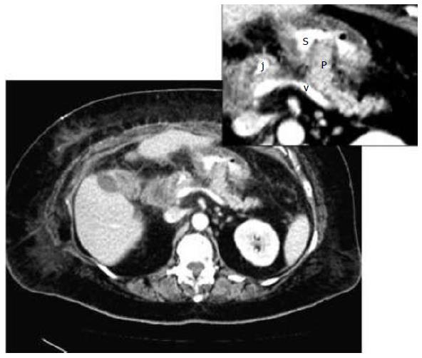
Figure 2 A scanner of the pancreaticogastrostomy in the early postoperative term.
In enlarged view. p: Pancreatic stump throw the gastric wall; s: Gastric lumen containing oral contrast media; v: Splenic vein draining to the portal vein on the right side of the patient; j: High density image corresponding to the staplers of the cutting edge of the jejuna limb used in the hepatico-jejunostomy.
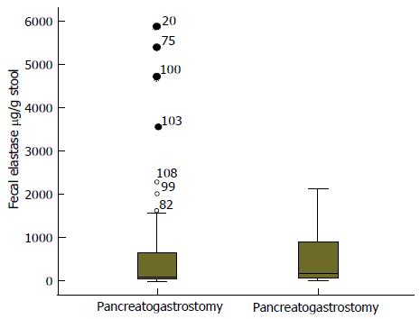
Figure 3 Fecal elastase levels in pancreatico-gastrostomy group and pancreatico-jejunostomy group.
Means are depicted with horizontal bars.
- Citation: Morera-Ocon FJ, Sabater-Orti L, Muñoz-Forner E, Pérez-Griera J, Ortega-Serrano J. Considerations on pancreatic exocrine function after pancreaticoduodenectomy. World J Gastrointest Oncol 2014; 6(9): 325-329
- URL: https://www.wjgnet.com/1948-5204/full/v6/i9/325.htm
- DOI: https://dx.doi.org/10.4251/wjgo.v6.i9.325


