Copyright
©2014 Baishideng Publishing Group Inc.
World J Gastrointest Oncol. Jul 15, 2014; 6(7): 194-210
Published online Jul 15, 2014. doi: 10.4251/wjgo.v6.i7.194
Published online Jul 15, 2014. doi: 10.4251/wjgo.v6.i7.194
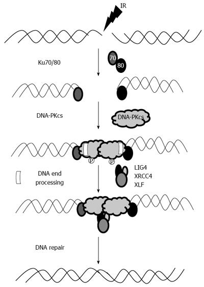
Figure 1 Schematic representations of double-strand break repair by non-homologous end-joining mechanism.
The KU proteins are the initial participants in this process as they rapidly bind to broken DNA segments. Another major function of the KU proteins is the active recruitment of DNA-PKcs. DNA-PK activation assists with the recruitment of other proteins involved in the limited DNAend-processing (Artemis, pol m, pol l, and TDK) required to generate ligatable DNA ends. Ligation is mediated by the LIG4/XRCC4 complex and is assisted by the ligation mediator XLF. Once this process is completed, DNA integrity is maintained.
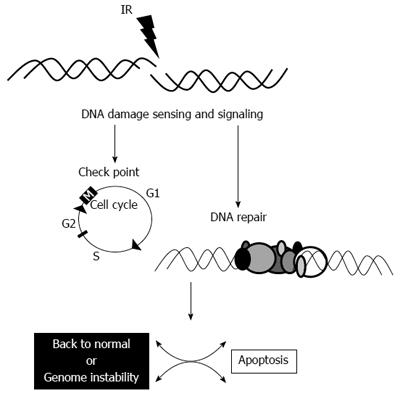
Figure 2 Schematic representation of the events that occur following IR-induced DNA damage.
Sensing mechanisms and signaling first stop the cell cycle that allows the cell to repair the DNA damage. If unsuccessful, apoptosis ensues. If the repair is nearly complete, the cell might continue to replicate with genome instability.
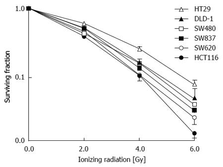
Figure 3 Response to ionizing radiation in several colorectal cancer cell lines subjected to various doses of ionizing radiation.
There is a variable response to the same doses or ionizing radiation (Gy).
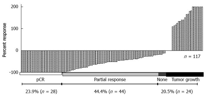
Figure 4 There is a high variability of a response to ionizing radiation in rectal cancer patients treated with pre-operative ionizing radiation.
Each bar on the X-axis represents an individual patient. The Y-axis represents the clinical response to pre-operative ionizing radiation. Nearly one fourth of patients achieve a pCR, but close to another fourth do not respond to the same form of treatment, while the rest of patients have achieve a partial response.
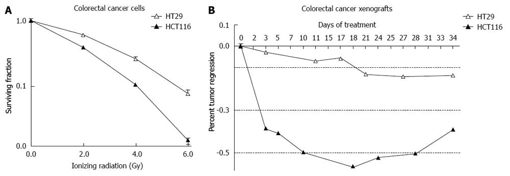
Figure 5 Analysis of the most radiosensitive (HCT116) and the most radioresistant (HT29) cells (A), a similar response has been noted in cells implanted in immune compromised mice bearing xenografts of these cells (B).
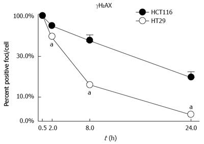
Figure 6 Analysis of DNA-induced damaged by ionizing radiation as determined by γH2AX.
HCT116 cells undergo more pronounced damage when treated with the same dose of ionizing radiation compared to HT29 cells. The damage induced in HCT116 cells persists over time. aP < 0.05 vs HCT116.
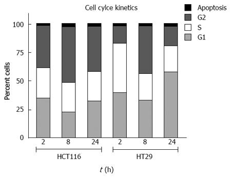
Figure 7 Cell cycle kinetics of HCT116 and HT29 cells treated with 2.
0 Gy ionizing radiation. There is a pronounced accumulation of cells in G2 in HCT116 cells. HT29 cells continue through the cell cycle in spite of receiving the same dose of ionizing radiation.
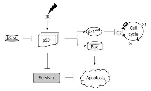
Figure 8 Schematic representation of molecular events following the cellular response to ionizing radiation-induced damage.
Ionizing radiation causes an up-regulation of p53. p53 then directly activates the cyclin dependent kinase inhibitor p21. Cell cycle progression stops until the cell repairs the damaged induced by ionizing radiation. If the cell is unable to repair itself, it undergoes apoptosis. Bcl-2 inhibits p53 up-regulation, while p53 inhibits the inhibitor of apoptosis: survivin.
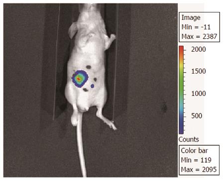
Figure 9 Orthotopic model for the study of rectal cancer.
This model has the following characteristics: (1) cecal transplantation of tumors with a known response to ionizing radiation; (2) attachment of the cecum to the lateral abdominal wall with a permanent suture for the administration of ionizing radiation; and (3) transfection of cells with luciferase before tumor implantation for the assessment of the chemoradiotherapeutic interventions over time by bioluminescence imaging before the end of the study. This technique allows targeted delivery of ionizing radiation in an intraperitoneal tumor.
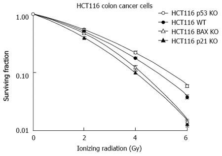
Figure 10 Analysis of HCT116 cells with stable KO genotypes for p53, p21, and Bax compared to wild-type.
Cells deficient in p53 are more radioresistant, while p21 and Bax deficient cells are more radiosensitive compared to wild-type.
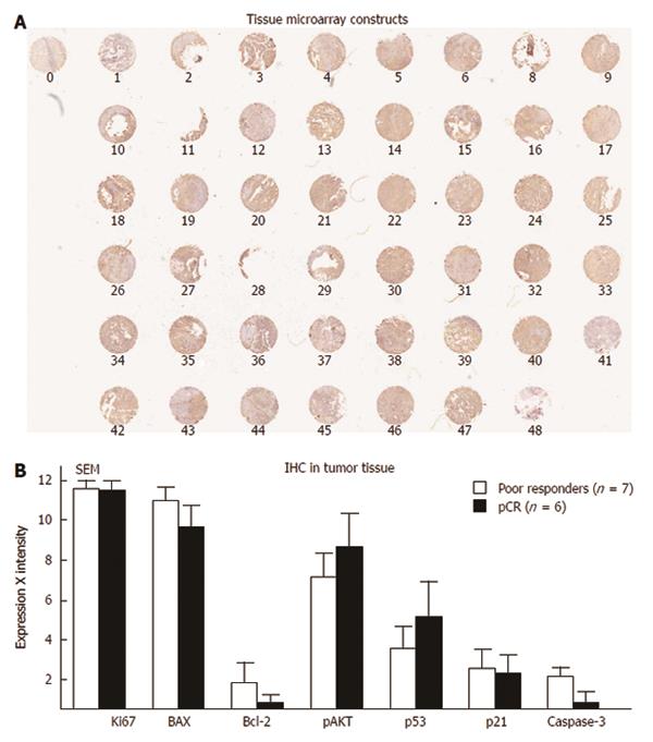
Figure 11 Tissue Microarray Constructs were created with 48 patients with rectal cancer that received pre-operative radiation.
A: Of these 48 patients (each dot represents a patient), six had a pCR and seven did not respond to treatment; B: The differences in various tumor markers comparing these two groups. IHC: Immunohistochemistry; SEM: Scanning electron microscope.
- Citation: Ramzan Z, Nassri AB, Huerta S. Genotypic characteristics of resistant tumors to pre-operative ionizing radiation in rectal cancer. World J Gastrointest Oncol 2014; 6(7): 194-210
- URL: https://www.wjgnet.com/1948-5204/full/v6/i7/194.htm
- DOI: https://dx.doi.org/10.4251/wjgo.v6.i7.194


