Copyright
©2012 Baishideng.
World J Gastrointest Oncol. May 15, 2012; 4(5): 119-124
Published online May 15, 2012. doi: 10.4251/wjgo.v4.i5.119
Published online May 15, 2012. doi: 10.4251/wjgo.v4.i5.119
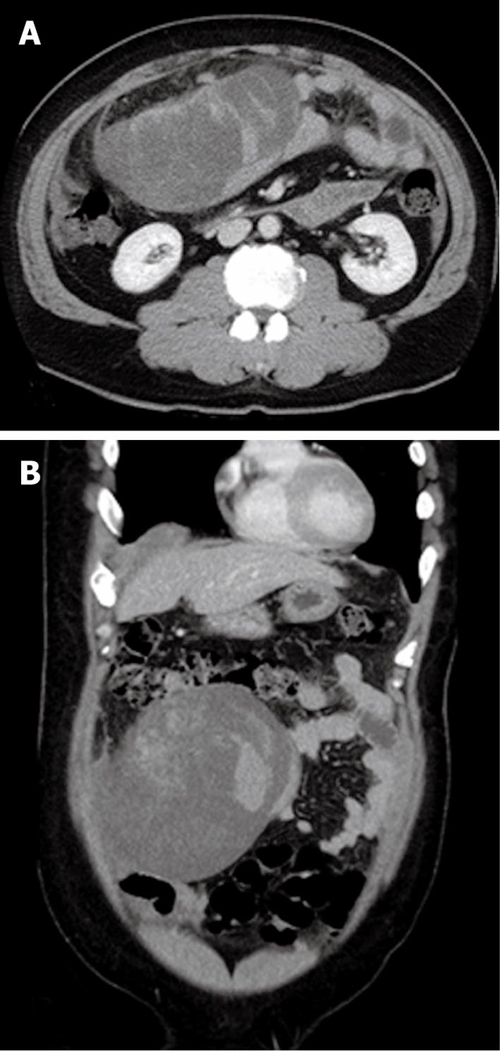
Figure 1 Contrast-enhanced axial (A) and coronal (B) abdominal computed tomography.
A huge mass was seen in the right abdominal cavity showing internal heterogeneity, with some high density area, but comprised mostly of low density regions which seemed to be a cystic component or interstitial mucous. The high density area was not enhanced, thus hemorrhage was suggested. Moreover a large amount of bloody ascites was seen in the pelvic cavity.
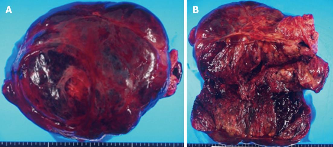
Figure 2 Gross appearance of the resected specimen.
A: The tumor appeared as a dark-red, relatively hard mass mixed with partly bleeding blood clots; B: The cut surface of the tumor showed cystic degeneration, and focal hemorrhage.
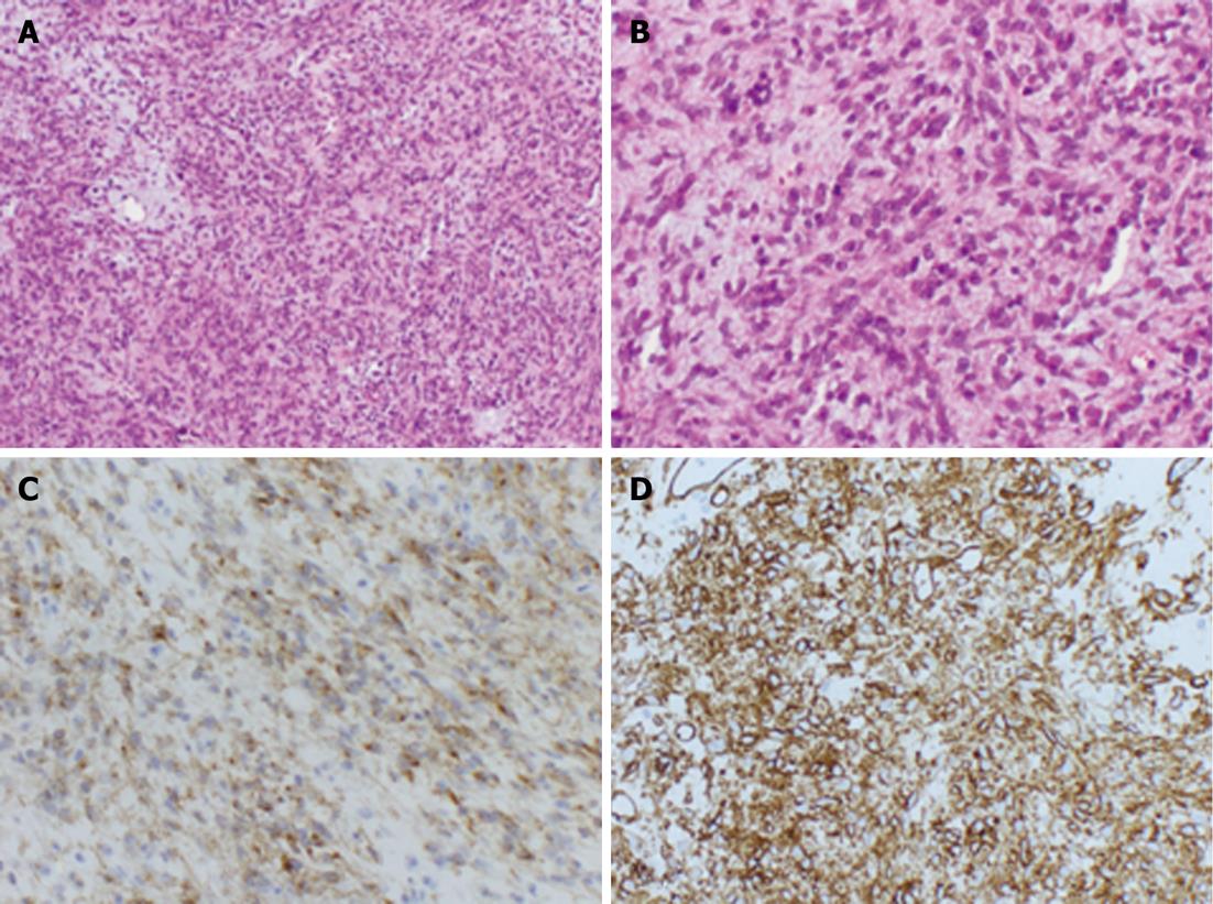
Figure 3 Histopathological findings of the resected specimen of the tumor.
A and B: The tumors were characterized by epithelioid-shaped cells and showed high cellularity (HE staining, A: × 40, B: × 200); C: Immunostaining of CD117 (KIT) was positive; D: Immunostaining of CD34 was positive (C, D: × 200).
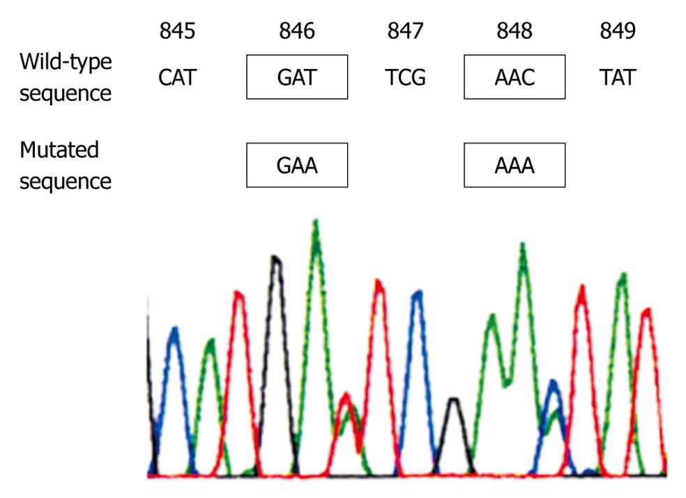
Figure 4 Representative sequencing results of omentum gastrointestinal stromal tumor.
This case showed a codon 846 (Asp to Glu) substitution, and a codon 848 (Asn to Lys) substitution within exon 18 of PDGFRA gene.
-
Citation: Murayama Y, Yamamoto M, Iwasaki R, Miyazaki T, Saji Y, Doi Y, Fukuda H, Hirota S, Hiratsuka M. Greater omentum gastrointestinal stromal tumor with
PDGFRA -mutation and hemoperitoneum. World J Gastrointest Oncol 2012; 4(5): 119-124 - URL: https://www.wjgnet.com/1948-5204/full/v4/i5/119.htm
- DOI: https://dx.doi.org/10.4251/wjgo.v4.i5.119


