Copyright
©2012 Baishideng.
World J Gastrointest Oncol. May 15, 2012; 4(5): 115-118
Published online May 15, 2012. doi: 10.4251/wjgo.v4.i5.115
Published online May 15, 2012. doi: 10.4251/wjgo.v4.i5.115
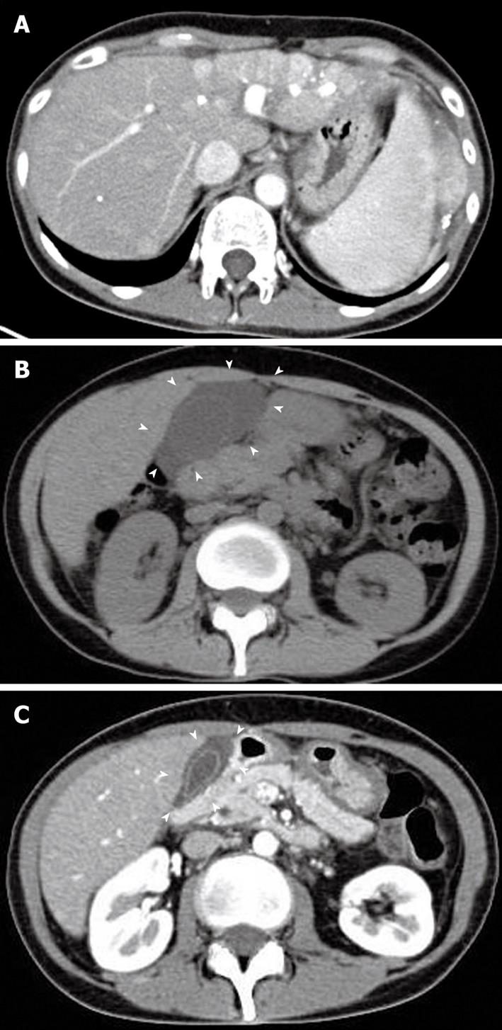
Figure 1 Abdominal computed tomography scan of the patient.
A: Enhanced abdominal computed tomography (CT) scan showing the diffuse HCC which occupied whole left lobe and several new lesions within diameter 1 cm in right liver before the treatment with sorafenib. Triangle indicates the HCC lesion; B: Abdominal CT scan at the onset of acute cholecystitis during the treatment with sorafenib. The CT revealed the gallbladder swelling with exudate associated with severe inflammation without stones or debris (arrowheads); C: Follow-up abdominal CT scan at 5 d after treatment of acute cholecystitis. Gallbladder swelling was markedly improved with reduced ascites exudate (arrowheads).
- Citation: Aihara Y, Yoshiji H, Yamazaki M, Ikenaka Y, Noguchi R, Morioka C, Kaji K, Tastumi H, Nakanishi K, Nakamura M, Yamao J, Toyohara M, Mitoro A, Sawai M, Yoshida M, Fujimoto M, Uemura M, Fukui H. A case of severe acalculous cholecystitis associated with sorafenib treatment for advanced hepatocellular carcinoma. World J Gastrointest Oncol 2012; 4(5): 115-118
- URL: https://www.wjgnet.com/1948-5204/full/v4/i5/115.htm
- DOI: https://dx.doi.org/10.4251/wjgo.v4.i5.115


