Copyright
©2011 Baishideng Publishing Group Co.
World J Gastrointest Oncol. May 15, 2011; 3(5): 75-78
Published online May 15, 2011. doi: 10.4251/wjgo.v3.i5.75
Published online May 15, 2011. doi: 10.4251/wjgo.v3.i5.75
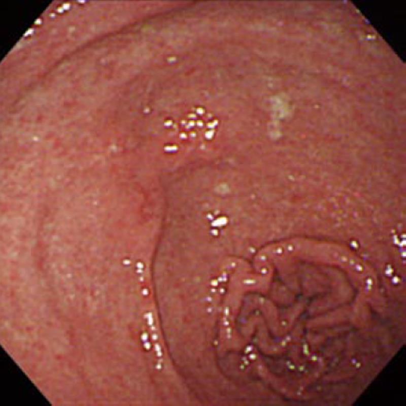
Figure 1 Endoscopic finding of gastric adenocarcinoma.
Gastrofiberscopy revealed an irregular depressed lesion with an obscure boundary in the lesser curvature of the prepyloric region.
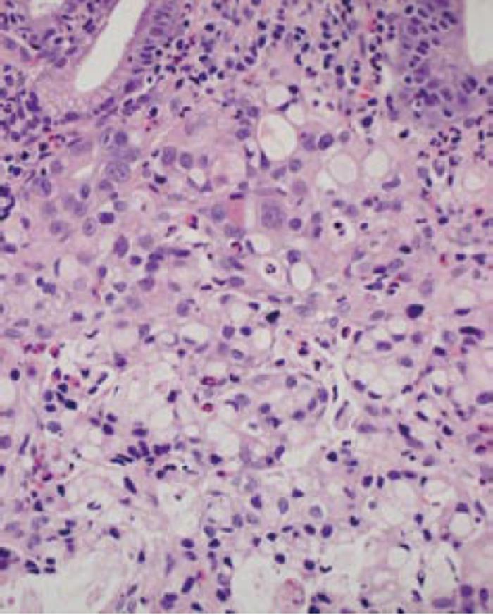
Figure 2 Histological finding of biopsy specimens.
Histological examination of biopsy specimens taken from the gastric lesion showed poorly differentiated adenocarcinoma that partially exhibited signet-ring cell morphology (Hematoxylin-and-eosin; original magnification, × 400).
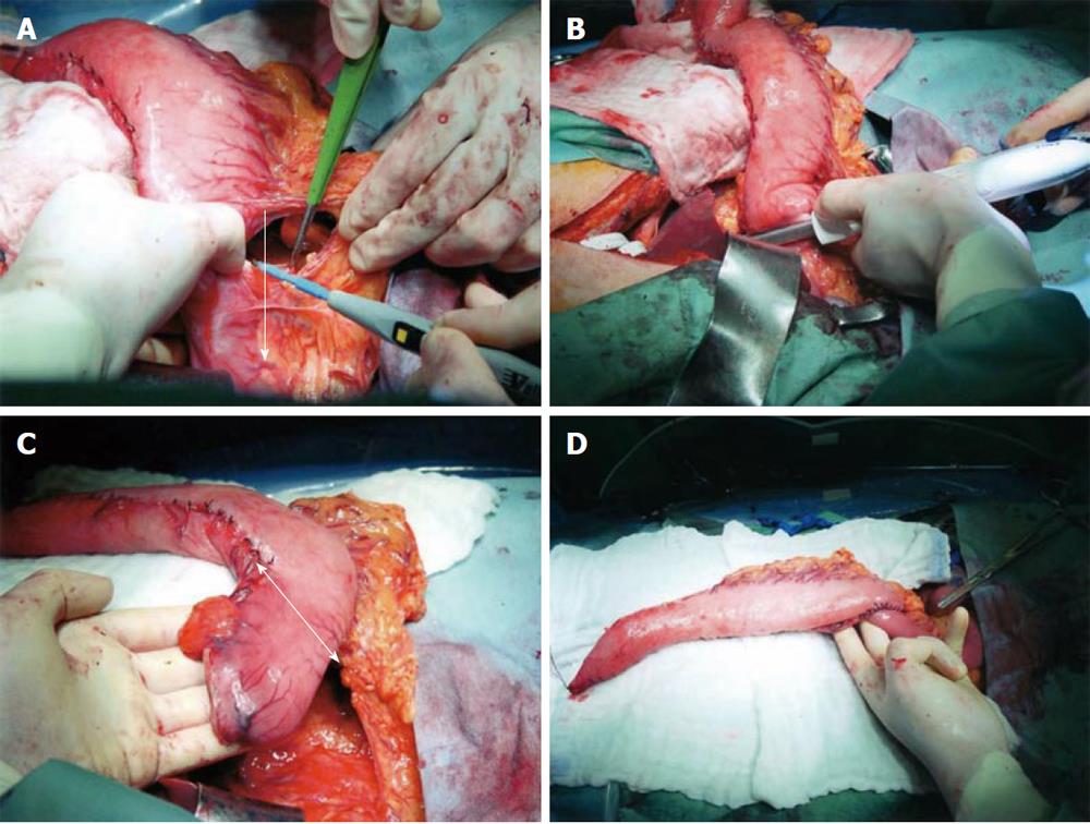
Figure 3 Intraoperative findings.
After a conventional gastric tube was prepared, the branches of the right gastroepiploic vessels supplying the pyloric lesion were cut along the greater curvature: The arrow line indicates the direction and range of devascularization (A). The duodenum was divided immediately distal to the pyloric ring and the duodenal stump was closed (B). The distal part of the gastric tube was resected to remove the early gastric adenocarcinoma of the prepyloric region and the suprapyloric lymph nodes: the arrow line indicates the cutting line for antrectomy (C). The distal stump of the gastric tube was anastomosed to the upper jejunum for a Roux-en-Y reconstruction prior to gastric pull-up to the neck (D).
- Citation: Kanda T, Sato Y, Yajima K, Kosugi SI, Matsuki A, Ishikawa T, Bamba T, Umezu H, Suzuki T, Hatakeyama K. Pedunculated gastric tube interposition in an esophageal cancer patient with prepyloric adenocarcinoma. World J Gastrointest Oncol 2011; 3(5): 75-78
- URL: https://www.wjgnet.com/1948-5204/full/v3/i5/75.htm
- DOI: https://dx.doi.org/10.4251/wjgo.v3.i5.75


