Copyright
©2011 Baishideng Publishing Group Co.
World J Gastrointest Oncol. Feb 15, 2011; 3(2): 19-23
Published online Feb 15, 2011. doi: 10.4251/wjgo.v3.i2.19
Published online Feb 15, 2011. doi: 10.4251/wjgo.v3.i2.19
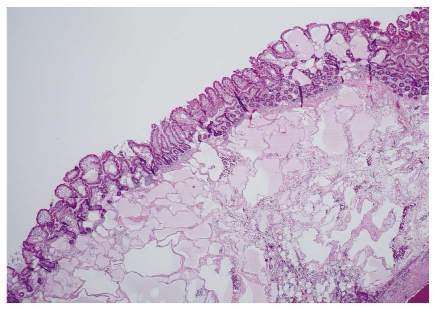
Figure 1 Low power view of dilated lymphatics involving mucosa and submucosa in intestinal lymphangiectasia.
In this section, the mucosal changes are patchy rather than continuous in distribution, emphasizing the multifocal nature of the disorder.
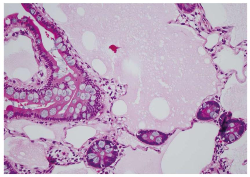
Figure 2 High power view showing mucosal involvement with dilated lymphatics in intestinal lymphangiectasia.
Surface epithelial cells are normal. Lymphatic protein is present in the dilated lymphatics of the intestinal mucosa.
- Citation: Freeman HJ, Nimmo M. Intestinal lymphangiectasia in adults. World J Gastrointest Oncol 2011; 3(2): 19-23
- URL: https://www.wjgnet.com/1948-5204/full/v3/i2/19.htm
- DOI: https://dx.doi.org/10.4251/wjgo.v3.i2.19


