Copyright
©2011 Baishideng Publishing Group Co.
World J Gastrointest Oncol. Oct 15, 2011; 3(10): 148-152
Published online Oct 15, 2011. doi: 10.4251/wjgo.v3.i10.148
Published online Oct 15, 2011. doi: 10.4251/wjgo.v3.i10.148
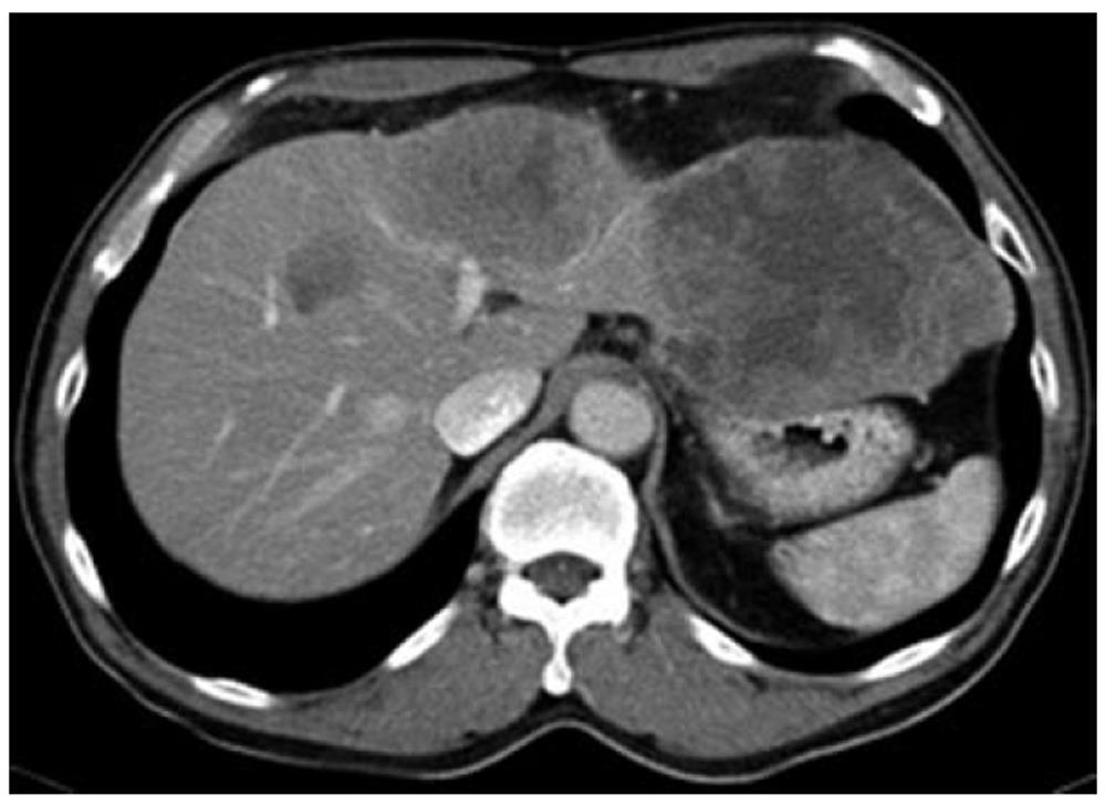
Figure 1 Computed tomography scan showing a large heterogeneously enhancing tumor in the left lobe of the liver.
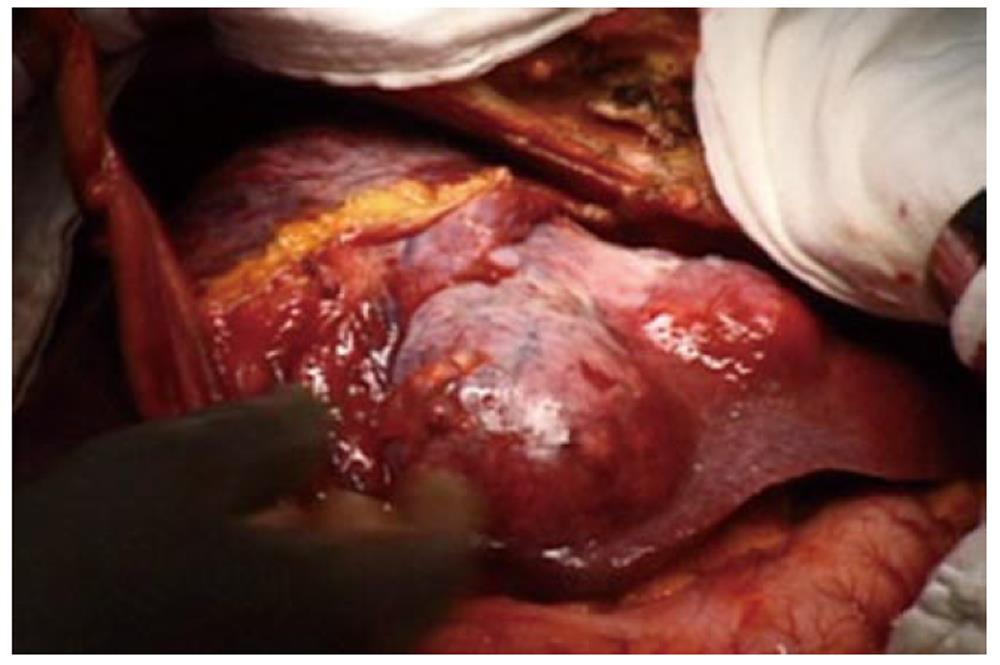
Figure 2 Intraoperative photo showing the large left lobe tumor.
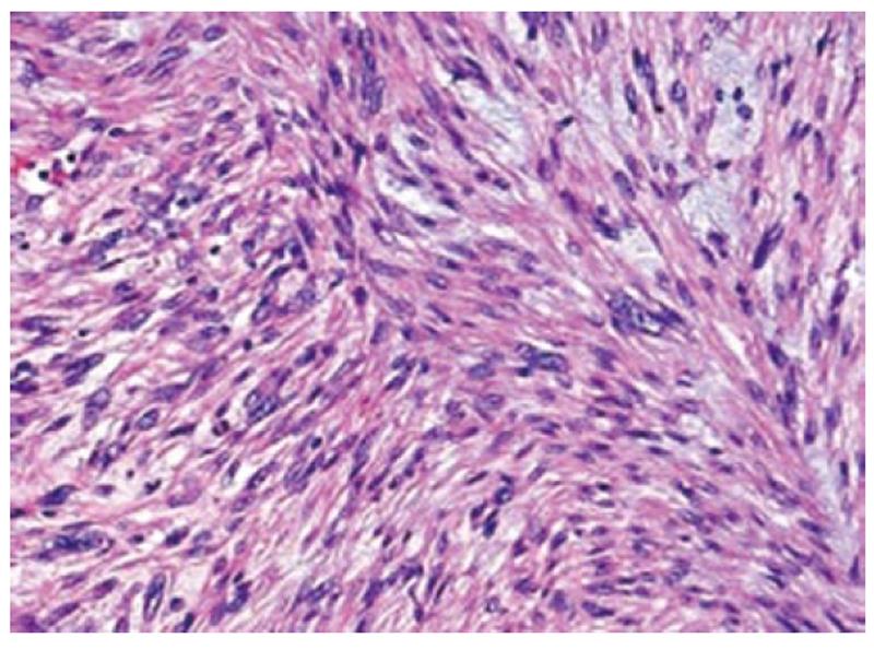
Figure 3 Histomicrograph showing fascicular growth pattern with tumor bundles intersecting each other at wide angles and merging of tumor cells with blood vessel walls.
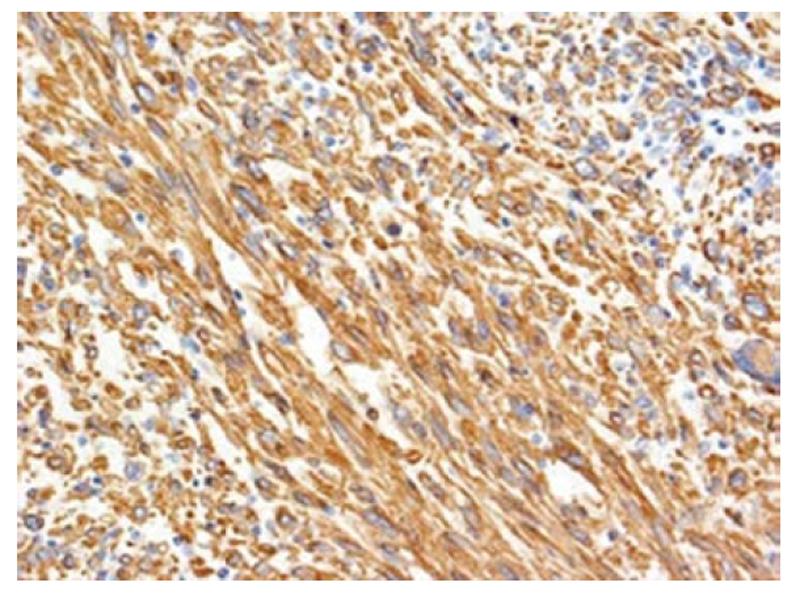
Figure 4 Tumor cells are positive for smooth muscle actin by immunohistochemical stain (× 100).
- Citation: Shivathirthan N, Kita J, Iso Y, Hachiya H, KyungHwa P, Sawada T, Kubota K. Primary hepatic leiomyosarcoma: Case report and literature review. World J Gastrointest Oncol 2011; 3(10): 148-152
- URL: https://www.wjgnet.com/1948-5204/full/v3/i10/148.htm
- DOI: https://dx.doi.org/10.4251/wjgo.v3.i10.148


