Copyright
©2010 Baishideng.
World J Gastrointest Oncol. Jun 15, 2010; 2(6): 282-286
Published online Jun 15, 2010. doi: 10.4251/wjgo.v2.i6.282
Published online Jun 15, 2010. doi: 10.4251/wjgo.v2.i6.282
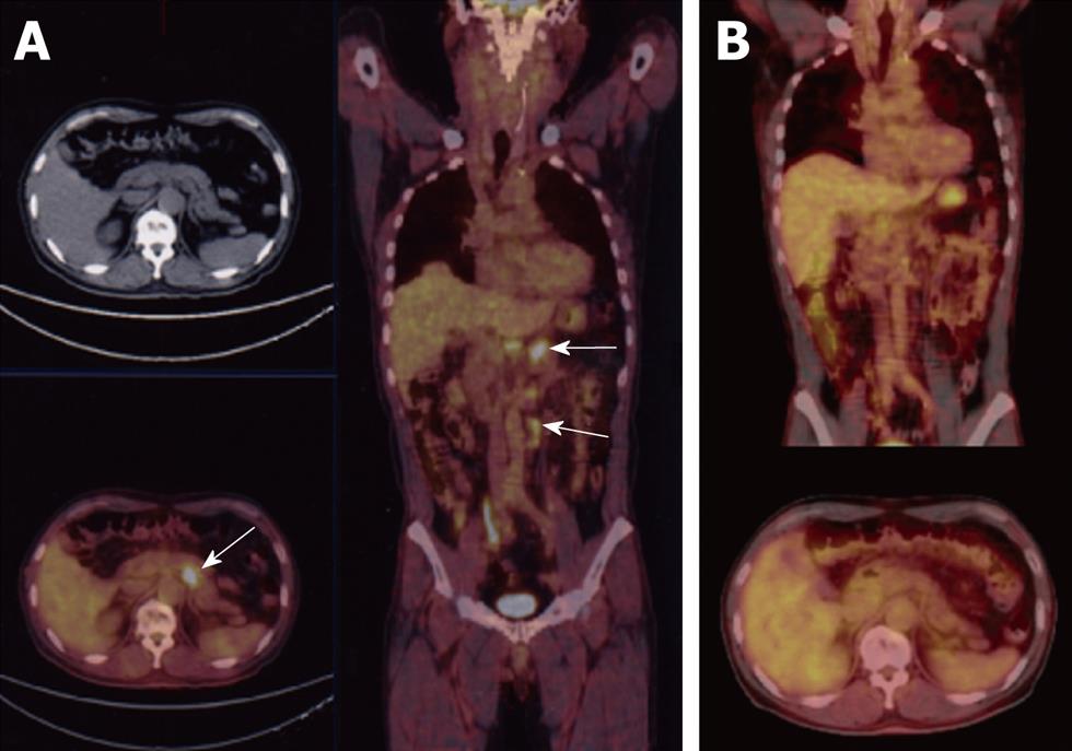
Figure 1 F-18 2-fluoro-2-deoxy-D-glucose (FDG) positron emission tomography (PET) computed tomography CT findings.
A: FDG-PET CT showing lymph nodes metastases in the paraaortic region (arrows); B: After chemoradiation therapy (CRT), FDG-PET CT demonstrated a marked reduction of the lymph nodes.
Figure 2 Gastrointestinal fiberscopy (GIF) and preoperative biopsy findings.
A: GIF before CRT demonstrating advanced type 2 gastric cancer at the antrum; B: Microscopic finding of the biopsy specimen obtained from the tumor, showing intestinal type adenocarcinoma (moderately differentiated tubular adenocarcinoma, Hematoxylin-Eosin 40X); C: GIF after CRT demonstrating the tiny erosion on the mucosa of antrum.
Figure 3 Abdominal CT scan findings.
A: CT scan showing lymph node metastasis in the lymph nodes around the celiac artery (arrow); B: Abdominal CT showing lymph node metastasis in the paraaortic region (arrow); C, D: After CRT, abdominal CT demonstrated a remarkable reduction of lymph node size.
Figure 4 Resected specimen and histopathological findings.
A, B: Macroscopic appearance of the surgically resected stomach. An ulcerative lesion was identified on the lesser curvature of the antrum. No tumor cells were observed in either the primary lesion (C, HE stain, 40×) or the dissected lymph nodes (D, HE stain, 40×), thus confirming a grade 3 effect (pathological complete response, pCR) for the treatment regimen.
- Citation: Shigeoka H, Imamoto H, Nishimura Y, Shimono T, Furukawa H, Imamura H, Yasuda T, Shiozaki H. Complete response to preoperative chemoradiotherapy in highly advanced gastric adenocarcinoma. World J Gastrointest Oncol 2010; 2(6): 282-286
- URL: https://www.wjgnet.com/1948-5204/full/v2/i6/282.htm
- DOI: https://dx.doi.org/10.4251/wjgo.v2.i6.282


