Copyright
©2010 Baishideng.
World J Gastrointest Oncol. Jun 15, 2010; 2(6): 272-281
Published online Jun 15, 2010. doi: 10.4251/wjgo.v2.i6.272
Published online Jun 15, 2010. doi: 10.4251/wjgo.v2.i6.272
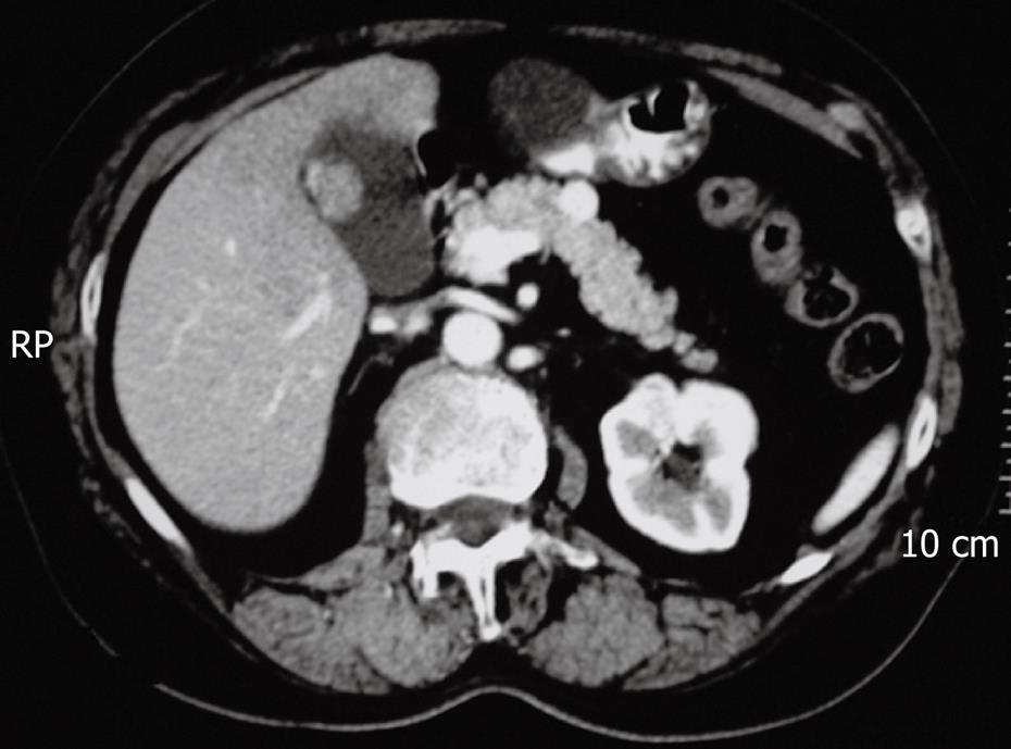
Figure 1 Contrast–enhanced tomography of the abdomen showing a neoplasm in the midportion of the pancreas (neuroendocrine tumor).
Figure 2 Surgical technique of central pancreatectomy[83].
A: Exposure of the anterior aspect of the pancreas and limits of resection; B: Transection of the pancreas including the lesion; C: Suture of the cephalic stump and partial dissection of splenic vessels; D: Anastomosis of the distal pancreatic stump with a jejunal loop.
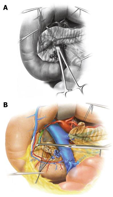
Figure 3 Duodenum-preserving pancreatic head resection (DPPHR)[84].
A: The superior mesenteric and the portal veins are cleared from the posterior aspect of the pancreas, and transection along the broken line is made; B: A rim of pancreatic tissue is left between the duodenal wall and the common bile duct that is skeletonized on the left side.
Figure 4 Residual head of the pancreas after (A) type-1, (B) type-2, and (C) type-3 DPPHR[84].
The residual rim of pancreatic tissue is located both superiorly and inferiorly to the major duodenal papilla of Vater in (A) type-1, only superiorly in (B) type-2, and totally removed in (C) type-3.
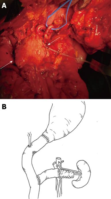
Figure 5 Middle-preserving pancreatectomy.
A: Intraoperative picture of remnant pancreas (arrows) after pylorus-preserving pancreaticoduodenectomy and pancreatic tail resection with preservation of splenic vessels and spleen; B: Final result of the operation with double jejunal loop reconstruction.
- Citation: Sperti C, Beltrame V, Milanetto AC, Moro M, Pedrazzoli S. Parenchyma-sparing pancreatectomies for benign or border-line tumors of the pancreas. World J Gastrointest Oncol 2010; 2(6): 272-281
- URL: https://www.wjgnet.com/1948-5204/full/v2/i6/272.htm
- DOI: https://dx.doi.org/10.4251/wjgo.v2.i6.272


