Copyright
©2010 Baishideng.
World J Gastrointest Oncol. Jun 15, 2010; 2(6): 251-258
Published online Jun 15, 2010. doi: 10.4251/wjgo.v2.i6.251
Published online Jun 15, 2010. doi: 10.4251/wjgo.v2.i6.251
Figure 1 Haematoxylin and eosin (H&E) stained sections showing the morphologic spectrum of goblet cell carcinoids (GCCs).
A: The typical cells that are encountered in GCCs: clusters or aggregates of cells with abundant mucin-filled cytoplasm that compresses the nucleus and resembles a goblet cell (× 100); B: In areas the tumor cells can be less conspicuous and they are separated by large amounts of fibrous stroma (× 200); C: In some cases, several of the tumor cells are found within pools of extravasated mucin (× 200); D: Extension of the tumor into periappendiceal or mesoappendiceal fat is a common finding (× 200).
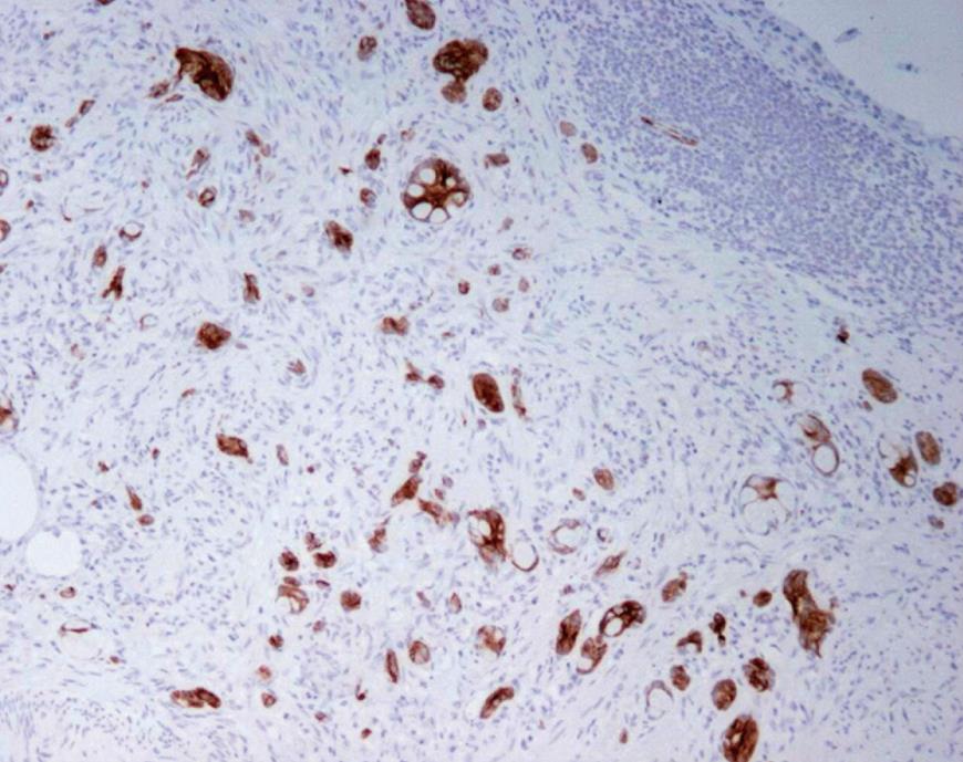
Figure 2 Tumor cells exhibit strong immunoreactivity for CK20 (× 200, anti-CK20).
- Citation: Roy P, Chetty R. Goblet cell carcinoid tumors of the appendix: An overview. World J Gastrointest Oncol 2010; 2(6): 251-258
- URL: https://www.wjgnet.com/1948-5204/full/v2/i6/251.htm
- DOI: https://dx.doi.org/10.4251/wjgo.v2.i6.251


