Copyright
©2010 Baishideng.
World J Gastrointest Oncol. May 15, 2010; 2(5): 247-250
Published online May 15, 2010. doi: 10.4251/wjgo.v2.i5.247
Published online May 15, 2010. doi: 10.4251/wjgo.v2.i5.247
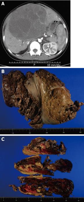
Figure 1 Computed tomography (CT) image and resected specimen.
A: Abdominal CT image before surgery; B: Resected specimen. The liver was ruptured; C: Cutting of the surface revealed a soft white mass with multilocular appearance, fibrous pseudocapsules, and massive hemorrhage.
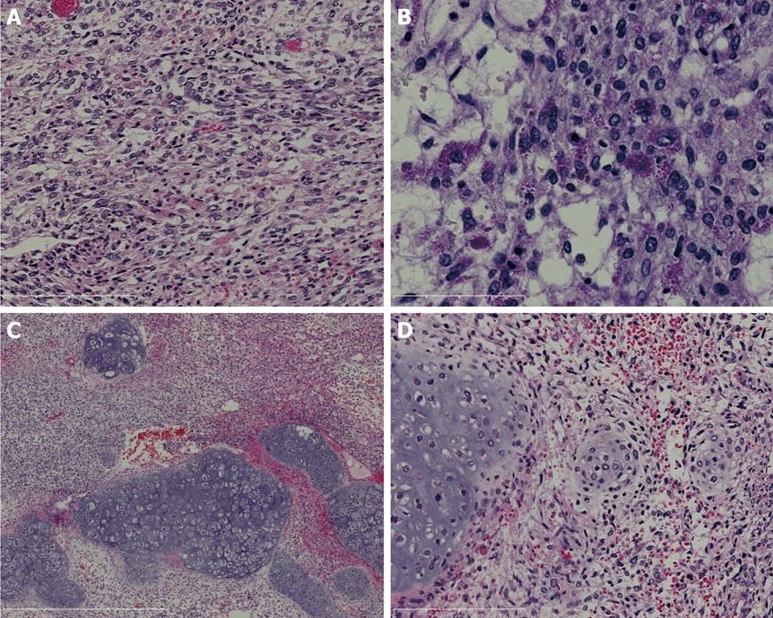
Figure 2 Histopathological findings.
A: Atypical spindle cells are proliferating and are arranged compactly or loosely (HE, bar = 200 μm); B: Some tumor cells contain eosinophilic hyaline globules of various sizes, which are resistant to diastase and positive for PAS (D-PAS, bar = 100 μm); C: In a small area, cartilage formation is observed (HE, bar = 1 mm); D: The cartilage component retains a lobulated appearance. The tumor cells exhibit nuclear atypia and anisonucleosis. Some cells are mitotic. As the cartilagenous matrix was deposited around the spindle tumor cells, tumor cells were seen to change to chondrosarcoma (HE, bar = 200 μm). HE: hematoxylin and eosin stain; D-PAS: Periodic acid-Schiff stain after diastase digestion.
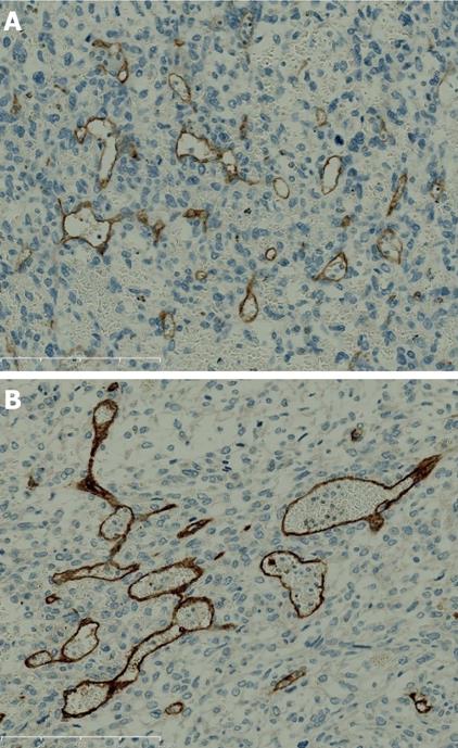
Figure 3 Immunohistochemical expression of ESL.
Tests for KIT (A) and CD34 (B) gave positive results for the endothelial cells in the tumor (bar = 200 μm). ESL; Embryonal sarcoma of the liver.
- Citation: Kinjo S, Sakurai S, Hirato J, Sunose Y. Embryonal sarcoma of the liver with chondroid differentiation. World J Gastrointest Oncol 2010; 2(5): 247-250
- URL: https://www.wjgnet.com/1948-5204/full/v2/i5/247.htm
- DOI: https://dx.doi.org/10.4251/wjgo.v2.i5.247


