Copyright
©2010 Baishideng.
World J Gastrointest Oncol. Mar 15, 2010; 2(3): 165-168
Published online Mar 15, 2010. doi: 10.4251/wjgo.v2.i3.165
Published online Mar 15, 2010. doi: 10.4251/wjgo.v2.i3.165
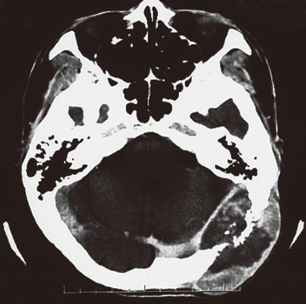
Figure 1 Contrast computed tomography (CT) showed hypervascular enhancement with osteolytic change in the skull.
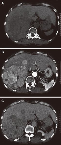
Figure 2 Contrast CT showed a huge enhanced mass in the liver.
A: Plain; B: Early phase; C: Late phase.
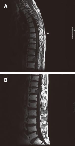
Figure 3 Magnetic resonance imaging showed bone metastasis in the thoracic vertebrae.
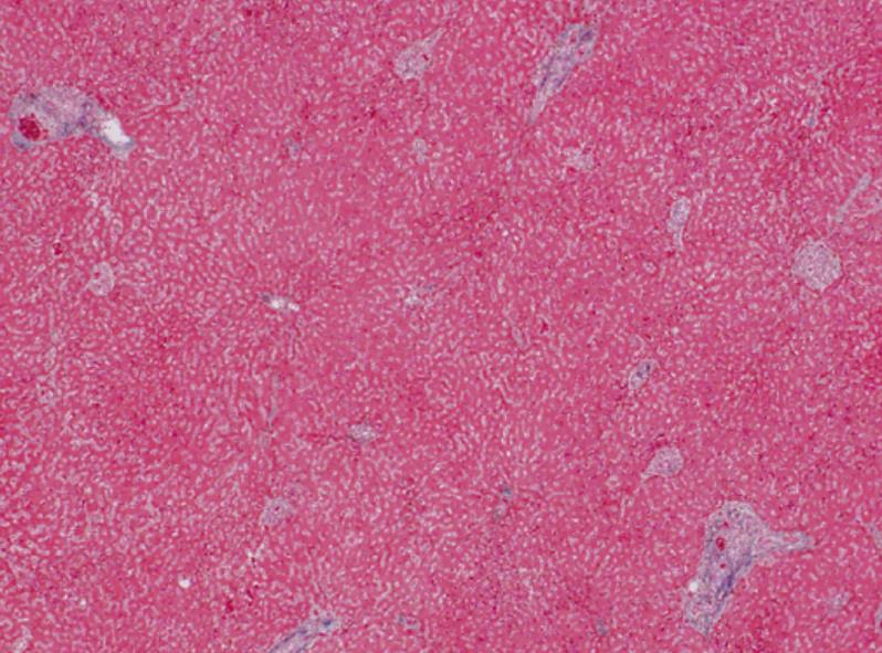
Figure 4 In the liver, mild infiltration of lymphocytes was seen in Glisson’s capsules which were significantly enlarged with well preserved limiting plates.
Piecemeal necrosis was not obvious. No fibrosis was noted.
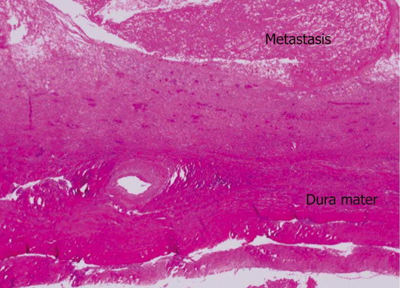
Figure 5 Dissection showed an 8 cm × 7 cm × 3 cm metastatic lesion had formed in the left occipitotemporal part of the cranial bone.
The lesion was osteolytic and showed invasion into the dura mater. Neither the subdural cavity nor the brain showed involvement from the metastatic tumor.
- Citation: Goto T, Dohmen T, Miura K, Ohshima S, Yoneyama K, Shibuya T, Kataoka E, Segawa D, Sato W, Anezaki Y, Ishii H, Kon D, Yamada I, Kamada K, Ohnishi H. Skull metastasis from hepatocellular carcinoma with chronic hepatitis B. World J Gastrointest Oncol 2010; 2(3): 165-168
- URL: https://www.wjgnet.com/1948-5204/full/v2/i3/165.htm
- DOI: https://dx.doi.org/10.4251/wjgo.v2.i3.165


