Copyright
©2010 Baishideng.
World J Gastrointest Oncol. Mar 15, 2010; 2(3): 146-150
Published online Mar 15, 2010. doi: 10.4251/wjgo.v2.i3.146
Published online Mar 15, 2010. doi: 10.4251/wjgo.v2.i3.146
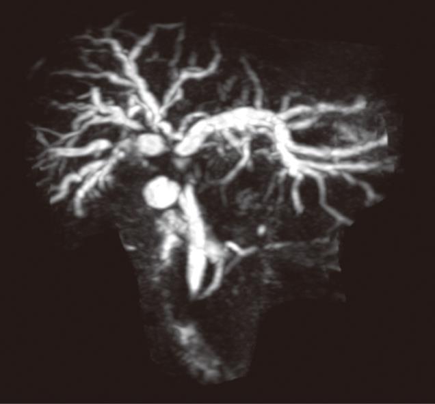
Figure 1 Magnetic resonance cholangiopancreatography demonstrating obstruction of the intrahepatic bile ducts due to a hilar cholangiocarcinoma.
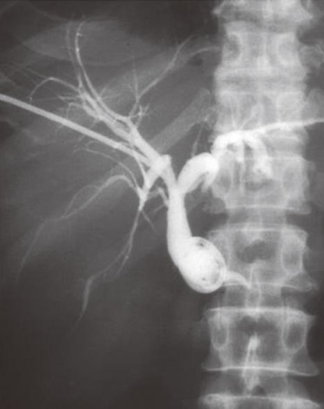
Figure 2 External biliary drainage catheter placed with common bile duct (CBD) obstruction secondary to pancreatic carcinoma.
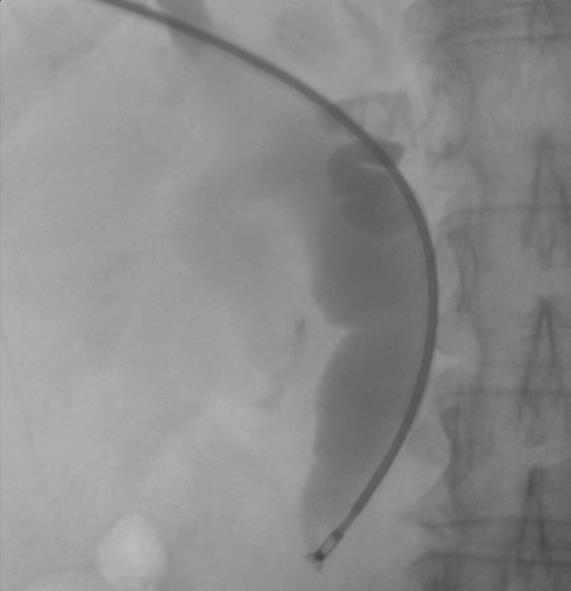
Figure 3 Biopsy forceps in use at the distal CBD in this patient with pancreatic carcinoma.
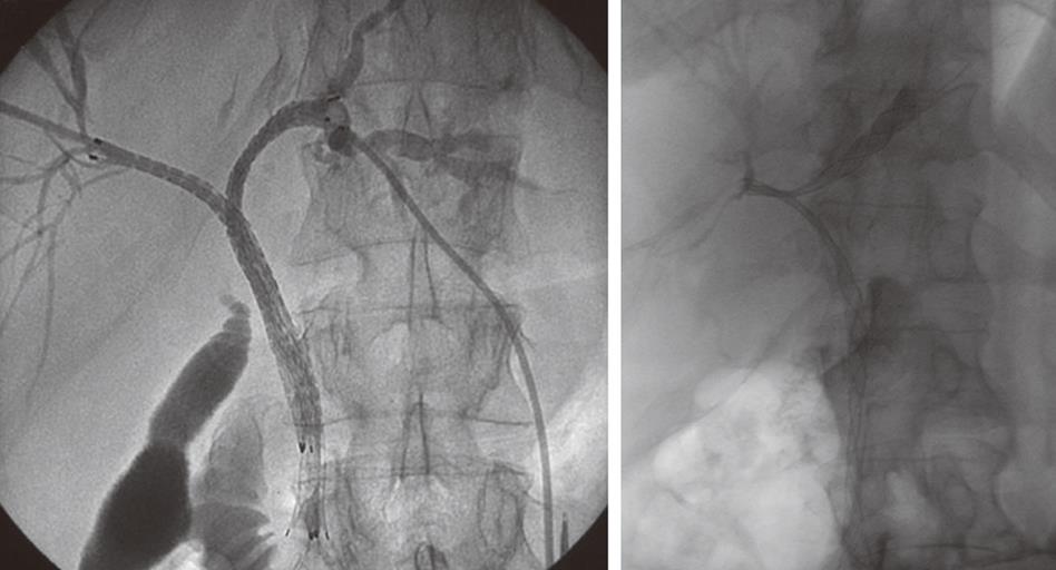
Figure 4 Images showing different configurations of stents for bi-lobar biliary drainage.
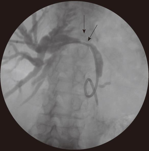
Figure 5 Internal-external drainage catheter in place across a small common hepatic duct cholangiocarcinoma (arrow).
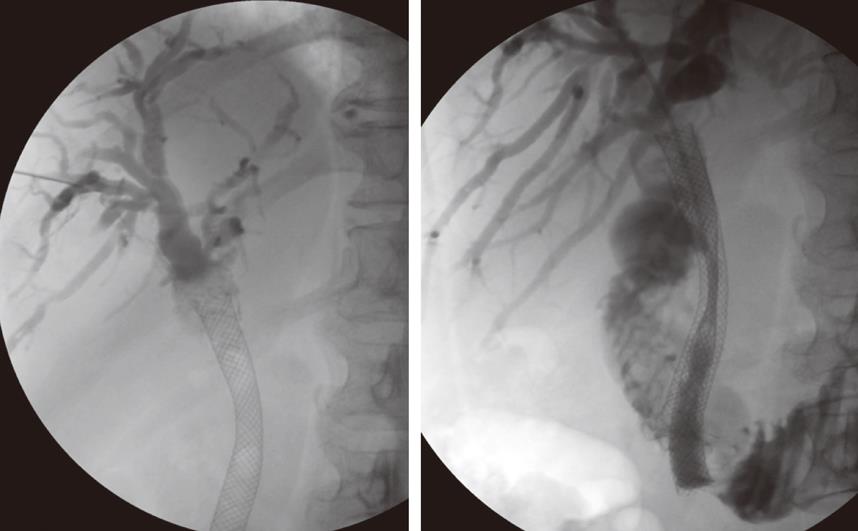
Figure 6 PTC shows tumor over-growth at the proximal end of the stent with no contrast flow distally.
Also, tumor in-growth as evidenced by lack of contrast opacification outside the new coaxially placed stent.
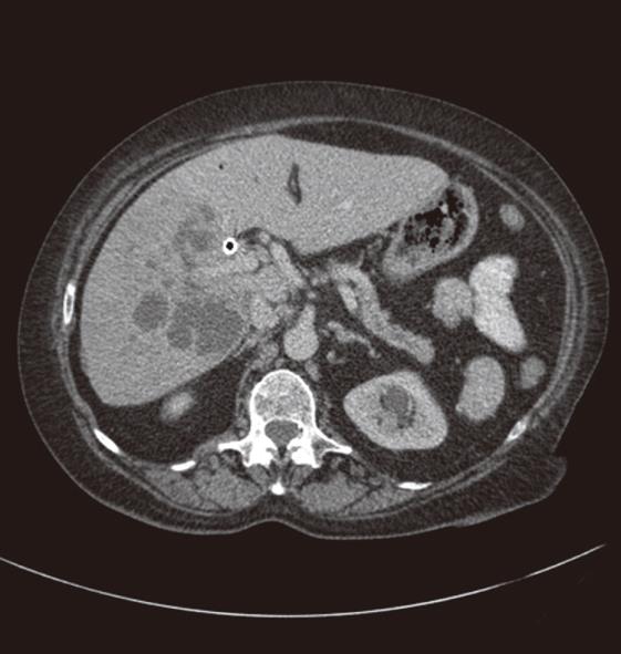
Figure 7 CT scan showing an abscess in the right lobe of liver, secondary to cholangitis affecting undrained right hepatic ducts.
- Citation: George C, Byass OR, Cast JEI. Interventional radiology in the management of malignant biliary obstruction. World J Gastrointest Oncol 2010; 2(3): 146-150
- URL: https://www.wjgnet.com/1948-5204/full/v2/i3/146.htm
- DOI: https://dx.doi.org/10.4251/wjgo.v2.i3.146


