Copyright
©2010 Baishideng.
World J Gastrointest Oncol. Jan 15, 2010; 2(1): 19-30
Published online Jan 15, 2010. doi: 10.4251/wjgo.v2.i1.19
Published online Jan 15, 2010. doi: 10.4251/wjgo.v2.i1.19
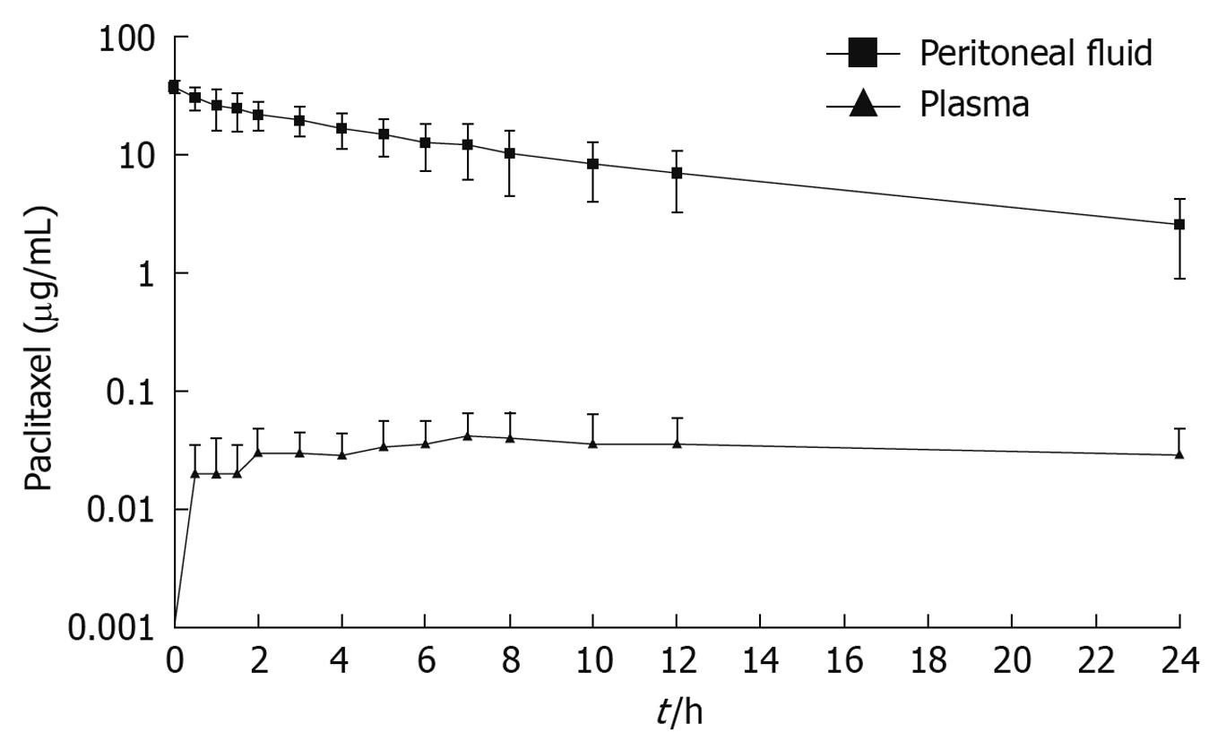
Figure 1 Pharmacokinetic study of concentration vs time for intraperitoneal paclitaxel.
The chemotherapy agent at 30 mg/m2 was instilled directly into the peritoneal cavity as rapidly as possible in a 1.5% dextrose peritoneal dialysis solution. The concentration of paclitaxel was determined in peritoneal fluid and in plasma for 24 h.
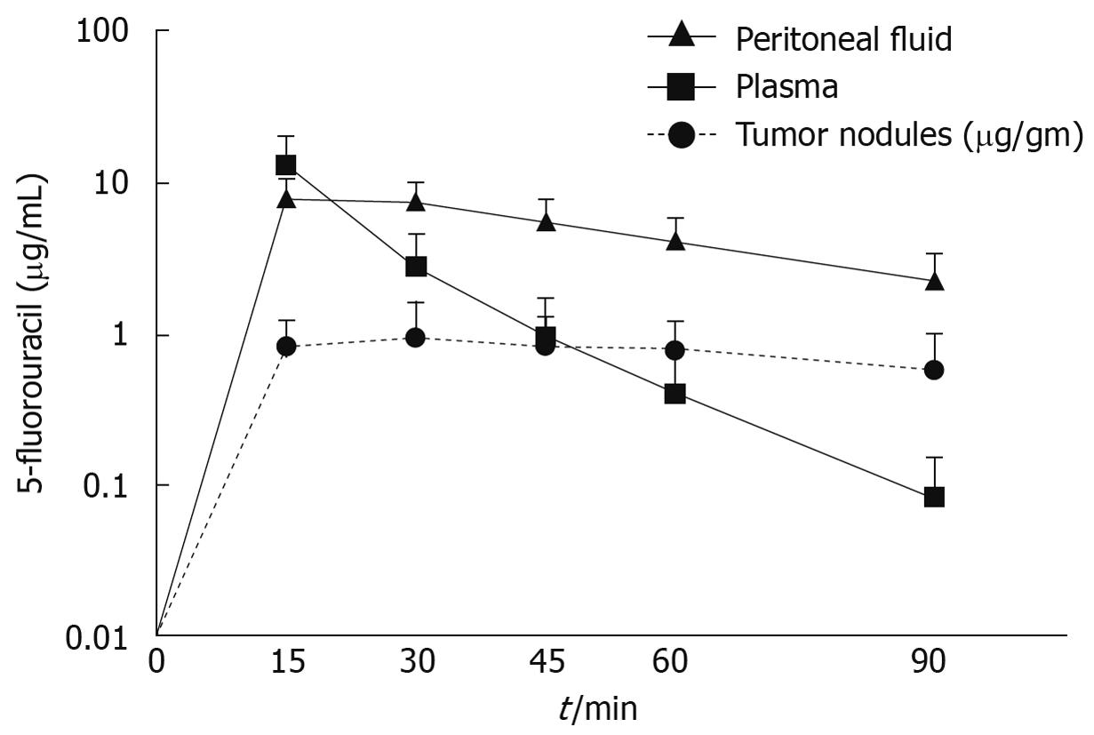
Figure 2 Pharmacodynamics during HIPEC after intravenous administration of 400 mg/m2 of 5-fluorouracil given simultaneously with intraperitoneal chemotherapy in 3 L of chemotherapy solution.
(Van der Speeten K, Stuart OA, Sugarbaker PH. Pharmacology of perioperative 5-fluorouracil. Submitted for publication).
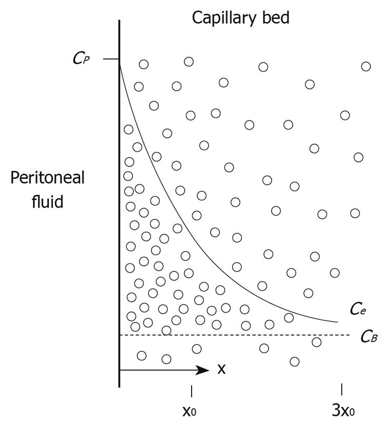
Figure 3 Conceptual diagram of tissue adjacent to the peritoneal cavity.
Cp = the free drug concentration in the peritoneal fluid; CB = the free drug concentration in the blood (or plasma). Solid line shows the exponential decrease in the free tissue interstitial concentration, Ce, as the drug diffuses down the concentration gradient and is removed by loss to the blood perfusing the tissue. Also shown are the characteristic diffusion length, x0, at which the concentration difference between the tissue and the blood has decreased to 37% of its maximum value, and 3x0, at which the difference has decreased to 5% of its maximum value[36].
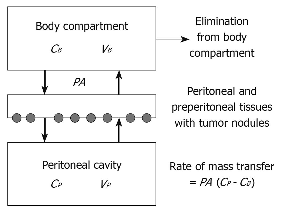
Figure 4 Three-compartment model of peritoneal transport, in which transfer of a drug from the peritoneal cavity to the blood occurs across the peritoneal membrane and preperitoneal tissues.
The peritoneal surface cancer nodules are located in these tissues. The permeability-area product (PA) governs this transfer and can be calculated by measuring the rate of drug disappearance from the cavity and dividing by the overall concentration difference between the peritoneal cavity and the blood (B). CB = the free drug concentration in the blood (or plasma); VB = volume of distribution of the drug in the body; CP = the free drug concentration in the peritoneal fluid; VP = volume of the peritoneal cavity[36].
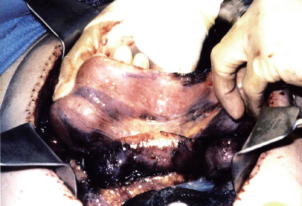
Figure 5 Spatial distribution of intraperitoneal methylene blue, using the closed abdomen technique.
Although the subcutaneous tissues were uniformly stained, the small bowel loops showed variable staining caused by adherence of adjacent bowel loops.
- Citation: Sugarbaker PH, Van der Speeten K, Stuart OA. Pharmacologic rationale for treatments of peritoneal surface malignancy from colorectal cancer. World J Gastrointest Oncol 2010; 2(1): 19-30
- URL: https://www.wjgnet.com/1948-5204/full/v2/i1/19.htm
- DOI: https://dx.doi.org/10.4251/wjgo.v2.i1.19


