Copyright
©The Author(s) 2025.
World J Gastrointest Oncol. Jun 15, 2025; 17(6): 106846
Published online Jun 15, 2025. doi: 10.4251/wjgo.v17.i6.106846
Published online Jun 15, 2025. doi: 10.4251/wjgo.v17.i6.106846
Figure 1 Moderate to large volumes of diffuse ascites in the abdominal and pelvic cavities, with smudged, nodular omentum extending from the epigastric region to the lower abdomen.
A: Coronal image; B: Peritoneal, seeding, nodular, mass lesions thickening in the cul-de-sac area, with focal abutment or invasion of the anterior wall of the mid-rectum; C: Sagittal view.
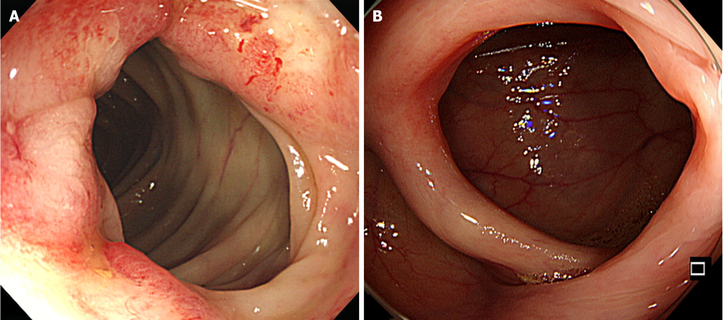
Figure 2 Colonoscopic features.
A: Ulcerofungating mass, 4 cm in diameter, was observed 45 cm from the anal verge during preoperative colonoscopy; B: Complete removal of the primary tumor was noted upon follow-up colonoscopy.
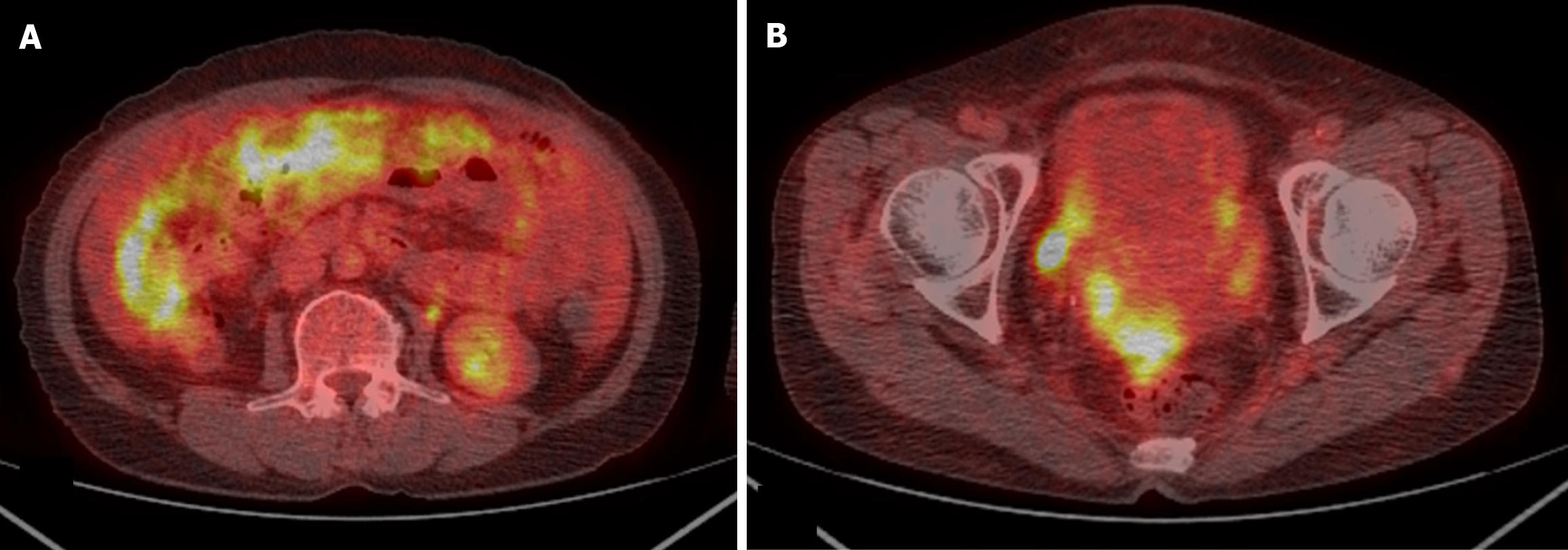
Figure 3 Positron emission tomography-computed tomography of the patient’s abdomen and pelvis.
A: Extensive hypermetabolic activity throughout the mesentery and omentum, suggesting diffuse peritoneal metastasis; B: Increased metabolic uptake in the pelvic region, indicating likely peritoneal seeding in the pelvic cavity.
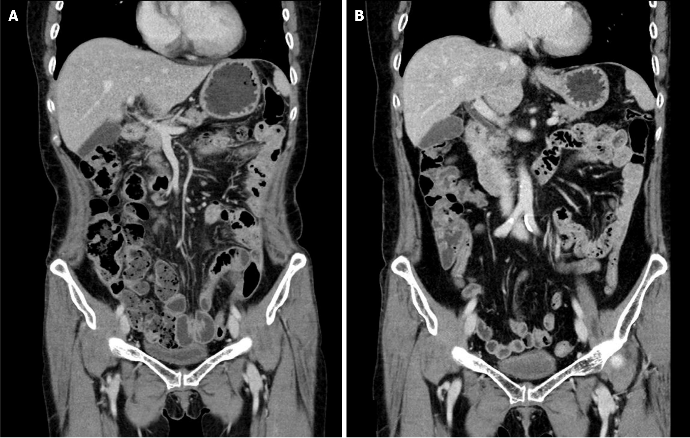
Figure 4 No sign of recurrence or metastasis is observed in either image.
A: Postoperative day 525; B: Postoperative day 2164.
- Citation: Suh JW, Jo JW, Park DG. Long-term survival of a patient with colorectal cancer with peritoneal carcinomatosis and low completeness of cytoreduction score: A case report. World J Gastrointest Oncol 2025; 17(6): 106846
- URL: https://www.wjgnet.com/1948-5204/full/v17/i6/106846.htm
- DOI: https://dx.doi.org/10.4251/wjgo.v17.i6.106846


