Copyright
©The Author(s) 2025.
World J Gastrointest Oncol. Mar 15, 2025; 17(3): 103328
Published online Mar 15, 2025. doi: 10.4251/wjgo.v17.i3.103328
Published online Mar 15, 2025. doi: 10.4251/wjgo.v17.i3.103328
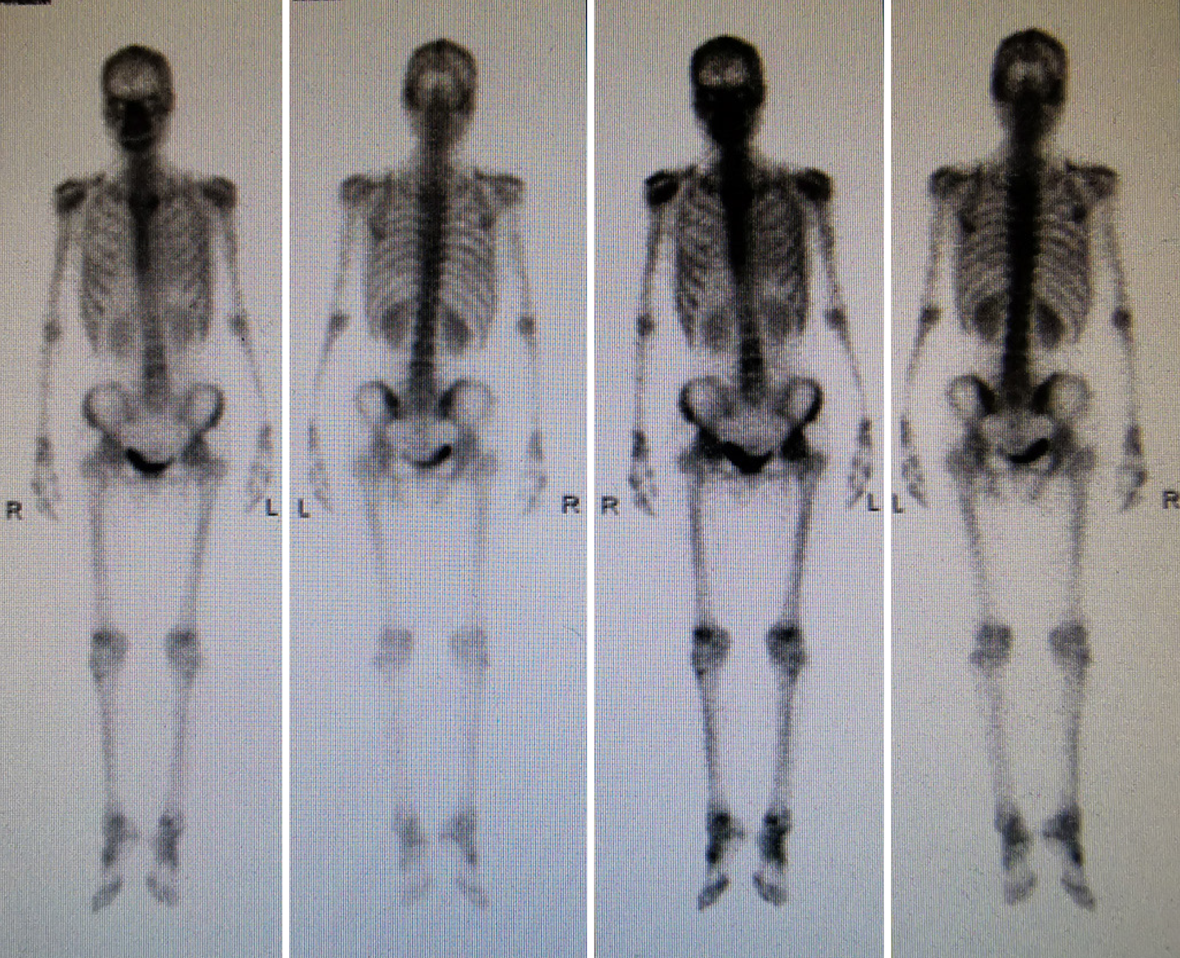
Figure 1 18F-fluorodeoxyglucose positron emission tomography/computed tomography.
18F-fluorodeoxyglucose (FDG) positron emission tomography/computed tomography images didn’t reveal high uptake of FDG in the gastrointestinal tract and other parts of the body in June 2021.
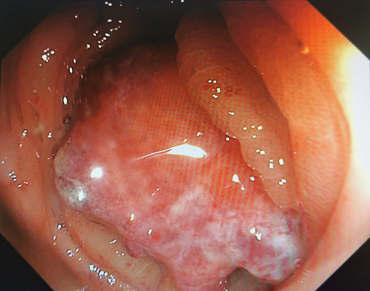
Figure 2 Colonoscopy showing a brittle mass in the ascending colon measuring about 4 cm × 5 cm in size.
The mass occupied the entire intestinal lumen. The lumen was narrow, and the colonoscope could not pass through it.
Figure 3 Contrast-enhanced computed tomography.
It showing the wall thickening of the ascending colon with a significantly enhanced soft tissue density mass causing an apparent stenosis (arrow).
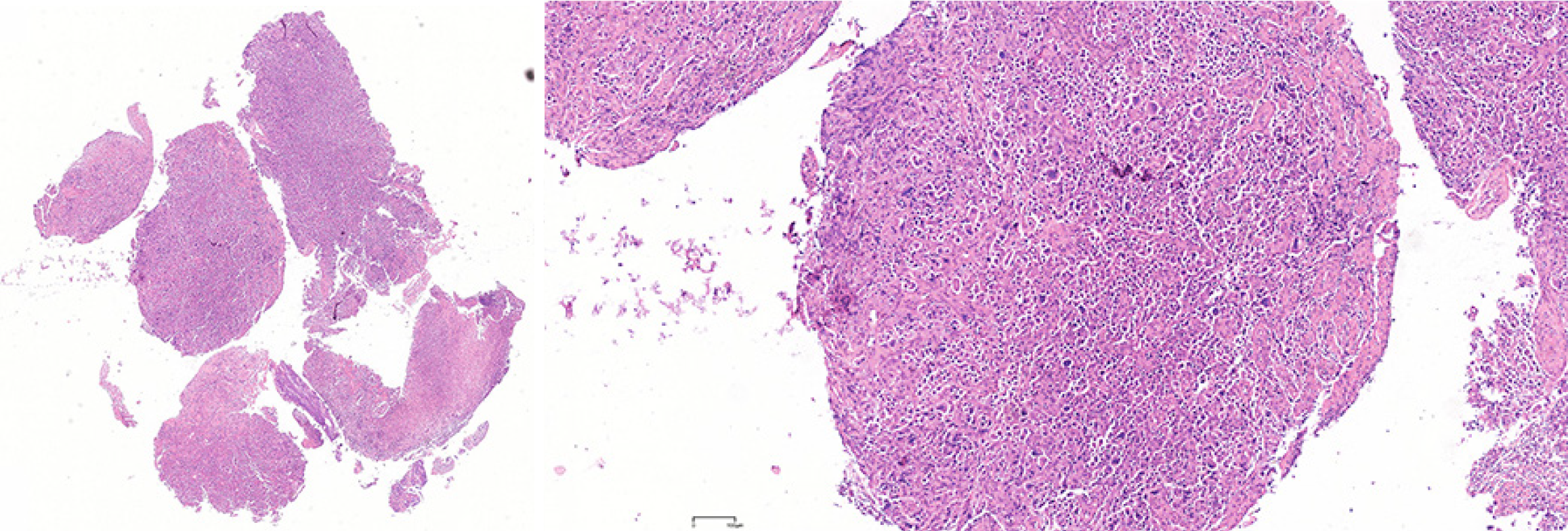
Figure 4 Colon biopsy.
It showing the diagnosis of poorly differentiated adenocarcinoma in the ascending colon.
Figure 5 Immunohistochemical staining of colon specimens.
A: Gross cystic disease fluid protein 15; B: GATA-binding protein 3; C: Mammaglobin; D: Cytokeratin 7. Tumor cells were positive.
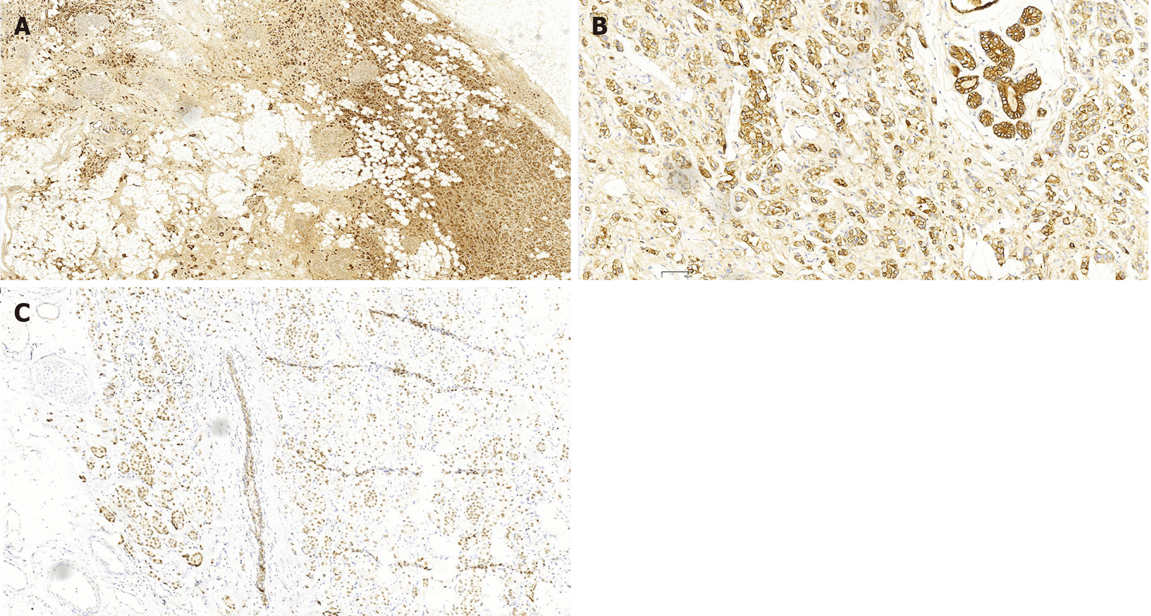
Figure 6 Immunohistochemical staining of breast cancer specimens.
A: Gross cystic disease fluid protein 15; B: GATA-binding protein 3; C: Cytokeratin 7. Tumor cells were positive.
- Citation: Wang BQ, Fang XG, Xiang BH, Wang XJ, Cao M, Zhuang CL, Liu ZC, Wang Z. Metastasizing to the colon from triple-negative breast cancer: A case report and review of literature. World J Gastrointest Oncol 2025; 17(3): 103328
- URL: https://www.wjgnet.com/1948-5204/full/v17/i3/103328.htm
- DOI: https://dx.doi.org/10.4251/wjgo.v17.i3.103328


