Copyright
©The Author(s) 2025.
World J Gastrointest Oncol. Feb 15, 2025; 17(2): 101780
Published online Feb 15, 2025. doi: 10.4251/wjgo.v17.i2.101780
Published online Feb 15, 2025. doi: 10.4251/wjgo.v17.i2.101780
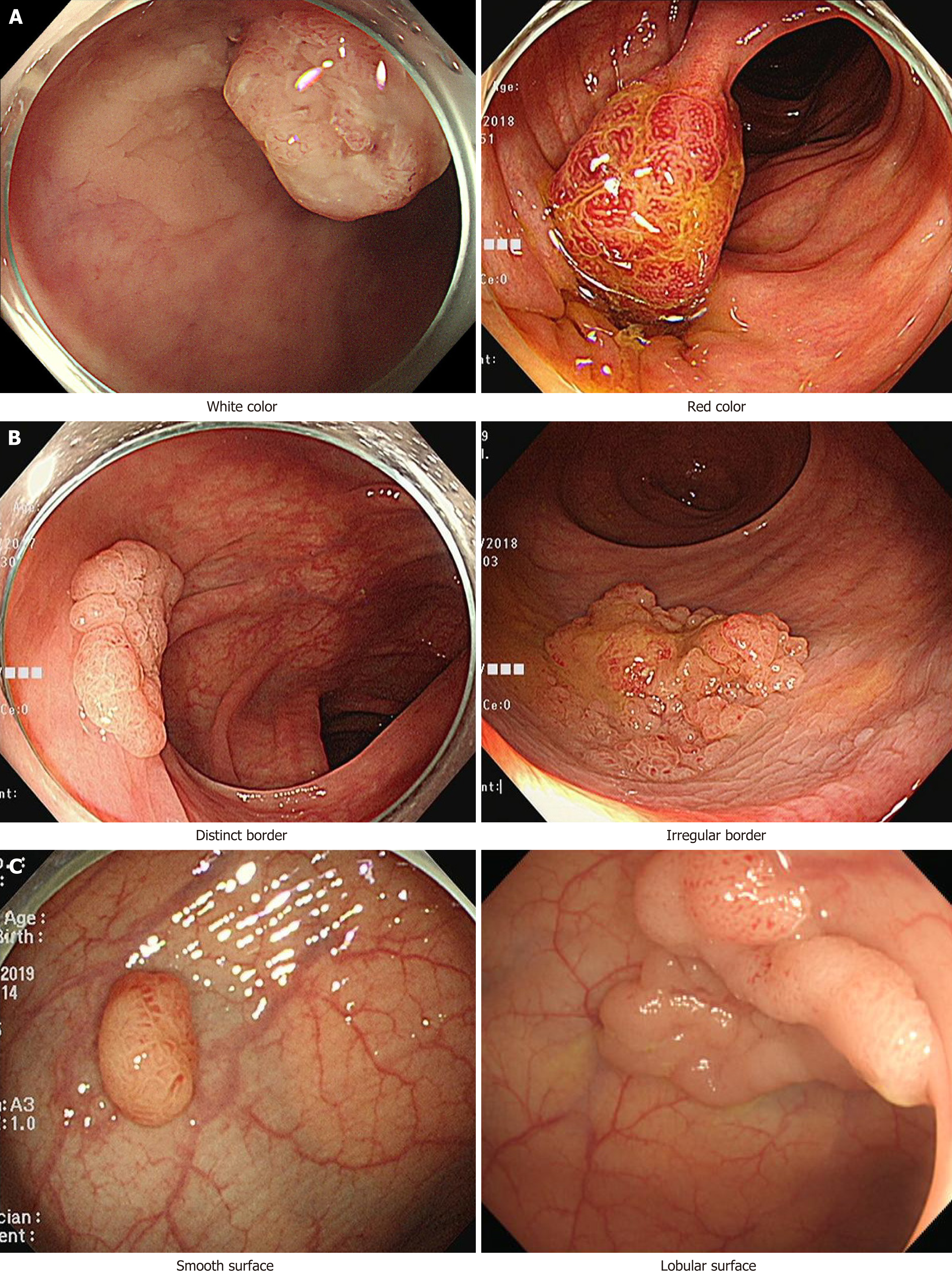
Figure 1 White-light colonoscopy images of traditional serrated adenomas in a representative case.
A: Color - white or red; B: Border - distinct or irregular; C: Surface - smooth or lobular.
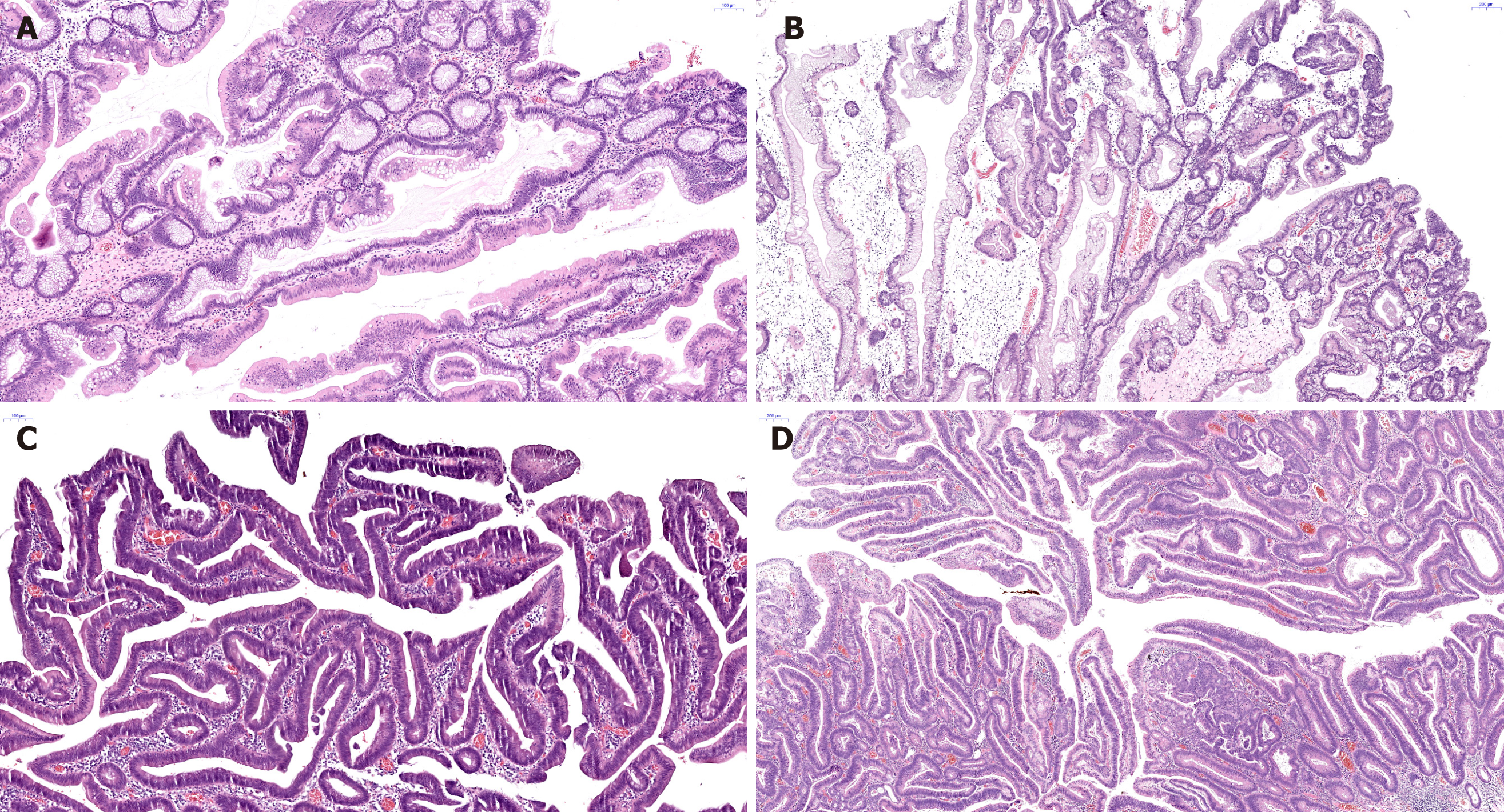
Figure 2 Histopathologic features observed with hematoxylin and eosin staining of the resected specimens of traditional serrated adenomas.
A: Traditional serrated adenomas (TSA) without dysplasia shows a saw-tooth appearance of the crypts with abundant eosinophilic cytoplasm and a basal/central elongated nucleus [hematoxylin and eosin (HE) staining, × 100]; B: TSA with low-grade dysplasia (HE staining, × 50); C: TSA with high-grade dysplasia (HE staining, × 100); D: TSA with adenocarcinoma (HE staining, × 50).
- Citation: Kim KH, Myung E, Oh HH, Im CM, Seo YE, Kim JS, Lim CJ, You GR, Cho SB, Lee WS, Noh MG, Lee KH, Joo YE. Clinical and endoscopic characteristics of colorectal traditional serrated adenomas with dysplasia/adenocarcinoma in a Korean population. World J Gastrointest Oncol 2025; 17(2): 101780
- URL: https://www.wjgnet.com/1948-5204/full/v17/i2/101780.htm
- DOI: https://dx.doi.org/10.4251/wjgo.v17.i2.101780


