Copyright
©The Author(s) 2025.
World J Gastrointest Oncol. Feb 15, 2025; 17(2): 100545
Published online Feb 15, 2025. doi: 10.4251/wjgo.v17.i2.100545
Published online Feb 15, 2025. doi: 10.4251/wjgo.v17.i2.100545
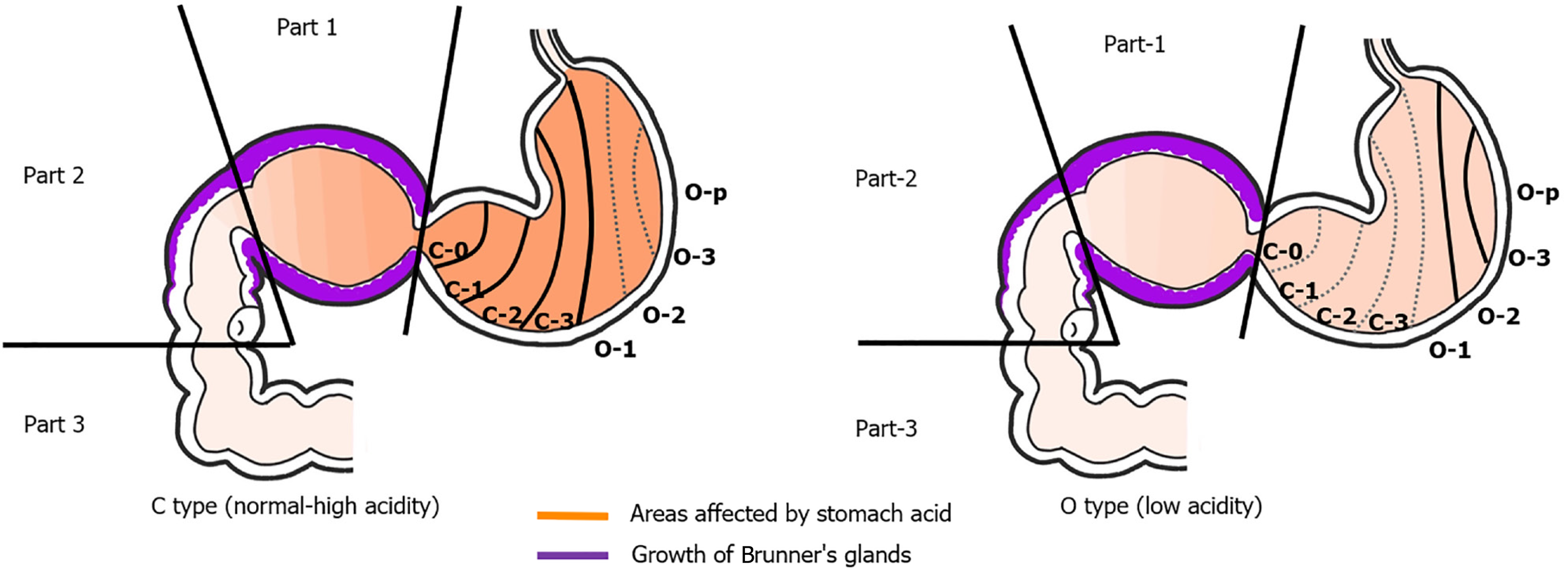
Figure 1 Endoscopic gastric mucosal atrophy according to the Kyoto classification of gastritis and effect of gastric acidity on the duodenum.
Part 1 comprises the bulb. Part 2 comprises the region from the superior duodenal angle to the papilla. Part 3 comprises the region from the anal side of the papilla to the horizontal part. Type C (C-0 to C-3) comprises no or mild endoscopic gastric mucosal atrophy. Type O (O-1 to O-p) comprises severe endoscopic gastric mucosal atrophy. Type C comprises normal to high acidity in the stomach and part 1. Type O comprises low acidity. Dark orange areas indicate highly acidic environments. Purple areas indicate the growth of Brunner’s glands.
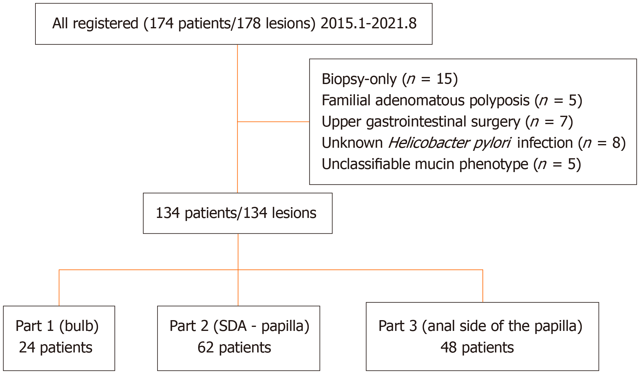
Figure 2 Patient flowchart.
SDA: Superior duodenal angle.
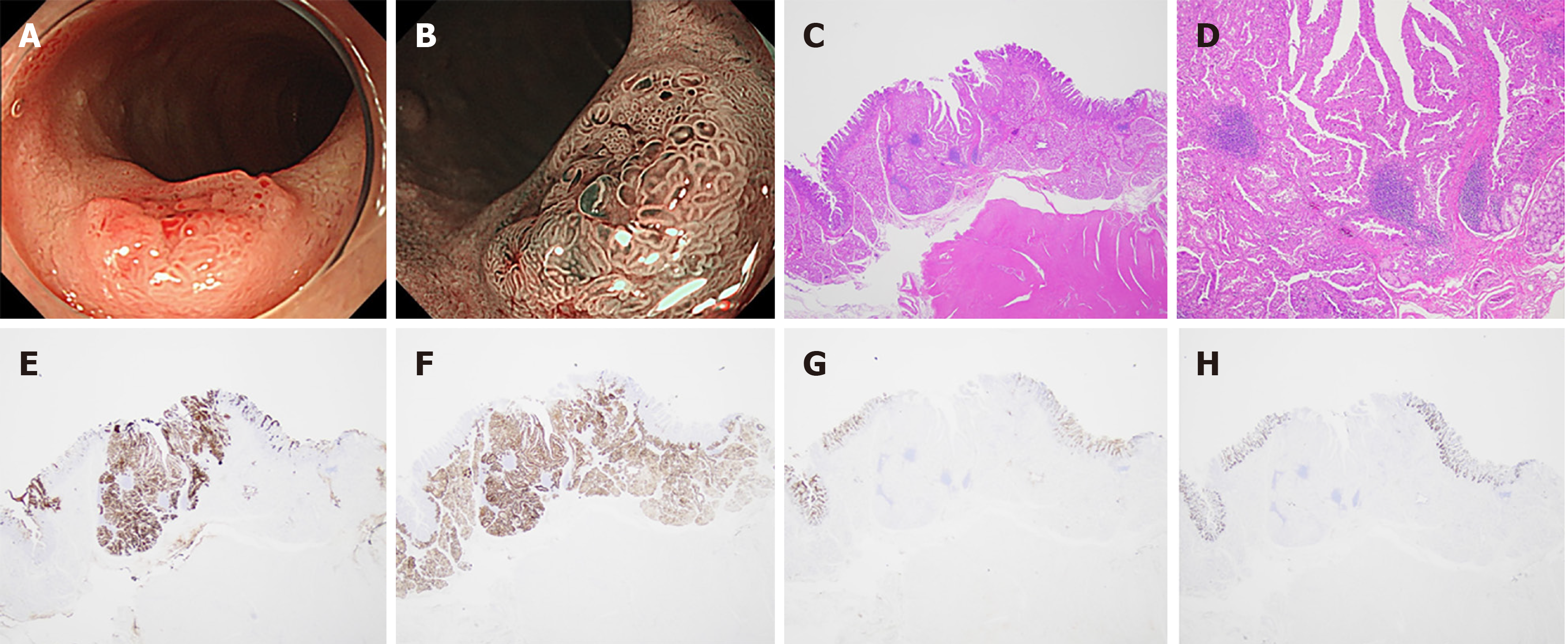
Figure 3 Non-ampullary duodenal epithelial tumors that express the gastric-type mucin phenotype.
A: White light imaging; B: Narrow band imaging; C: Hematoxylin-eosin stain; D: Hematoxylin-eosin stain (3000 μm invasion into the submucosa); E: MAC5AC; F: MAC6; G: CD10; H: MAC2.
- Citation: Ohno K, Nakatani E, Kurokami T, Kawai A, Itai R, Matsuda M, Masui Y, Satoh T, Ikeda S, Hirata T, Takeda S, Suzuki M, Haruma K. Relationship between gastric mucosal atrophy by endoscopy and non-ampullary duodenal epithelial tumors. World J Gastrointest Oncol 2025; 17(2): 100545
- URL: https://www.wjgnet.com/1948-5204/full/v17/i2/100545.htm
- DOI: https://dx.doi.org/10.4251/wjgo.v17.i2.100545


