Copyright
©The Author(s) 2024.
World J Gastrointest Oncol. Sep 15, 2024; 16(9): 4028-4036
Published online Sep 15, 2024. doi: 10.4251/wjgo.v16.i9.4028
Published online Sep 15, 2024. doi: 10.4251/wjgo.v16.i9.4028
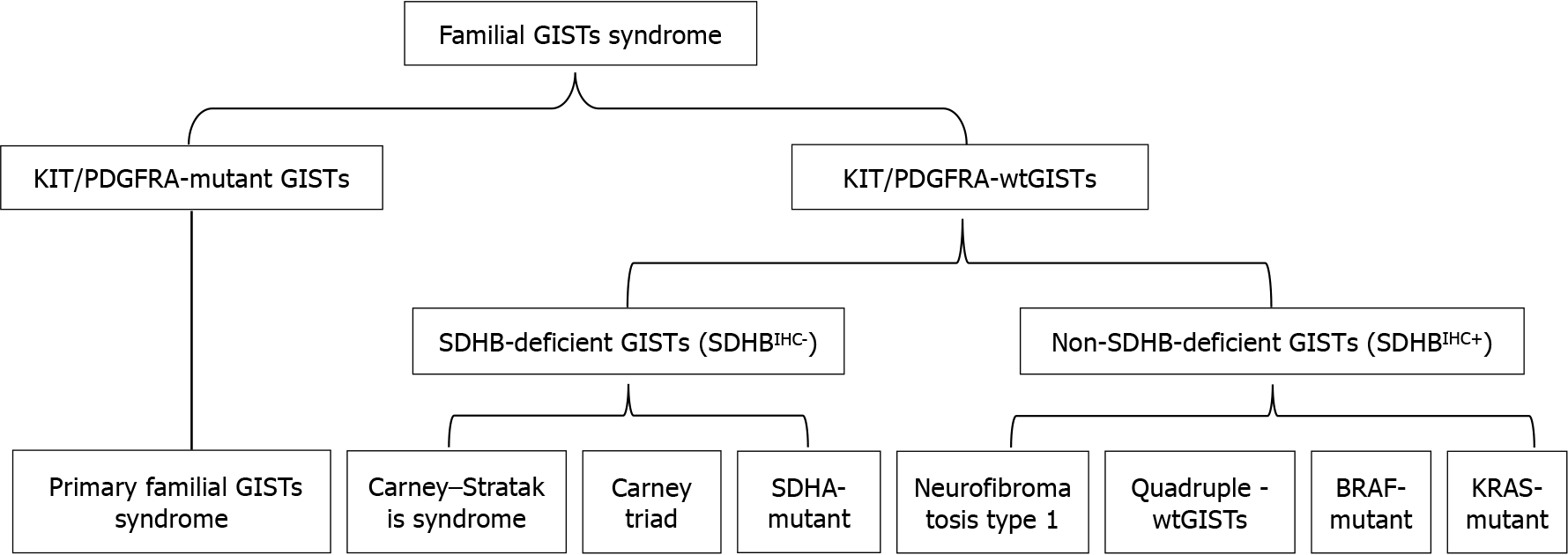
Figure 1 Familial gastrointestinal stromal tumor classification.
GISTs: Gastrointestinal stromal tumors; wtGISTs: Wild-type Gastrointestinal stromal tumors; SDHB: Succinate dehydrogenase complex subunit B; IHC: Immunohistochemical staining.
Figure 2 The family pedigree.
Square: Male; round: Female; GISTs: Gastrointestinal stromal tumors; wtGISTs: Wild-type Gastrointestinal stromal tumors.
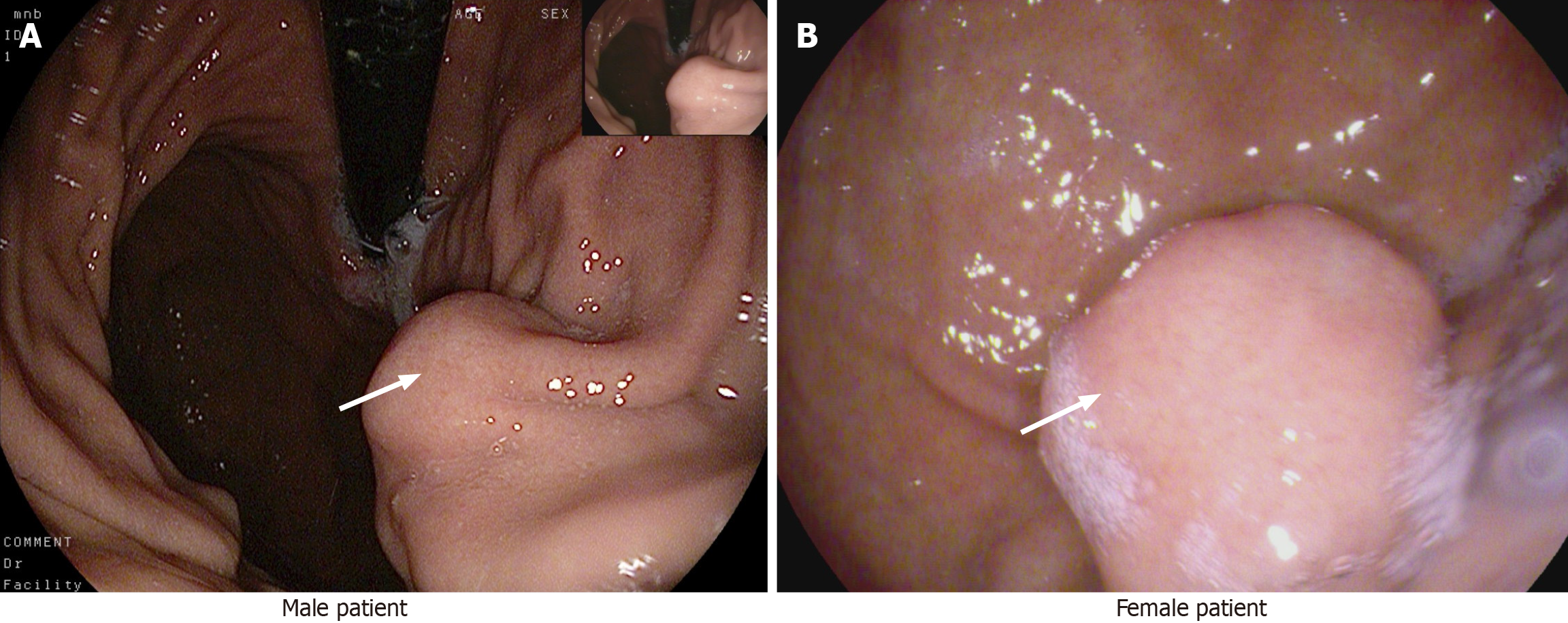
Figure 3 Preoperative gastroscopy results of both patients.
A: A submucosal lesion in the stomach body; B: A mucosal lesion approximately 1.5 cm × 2.0 cm in the fundus of the stomach.
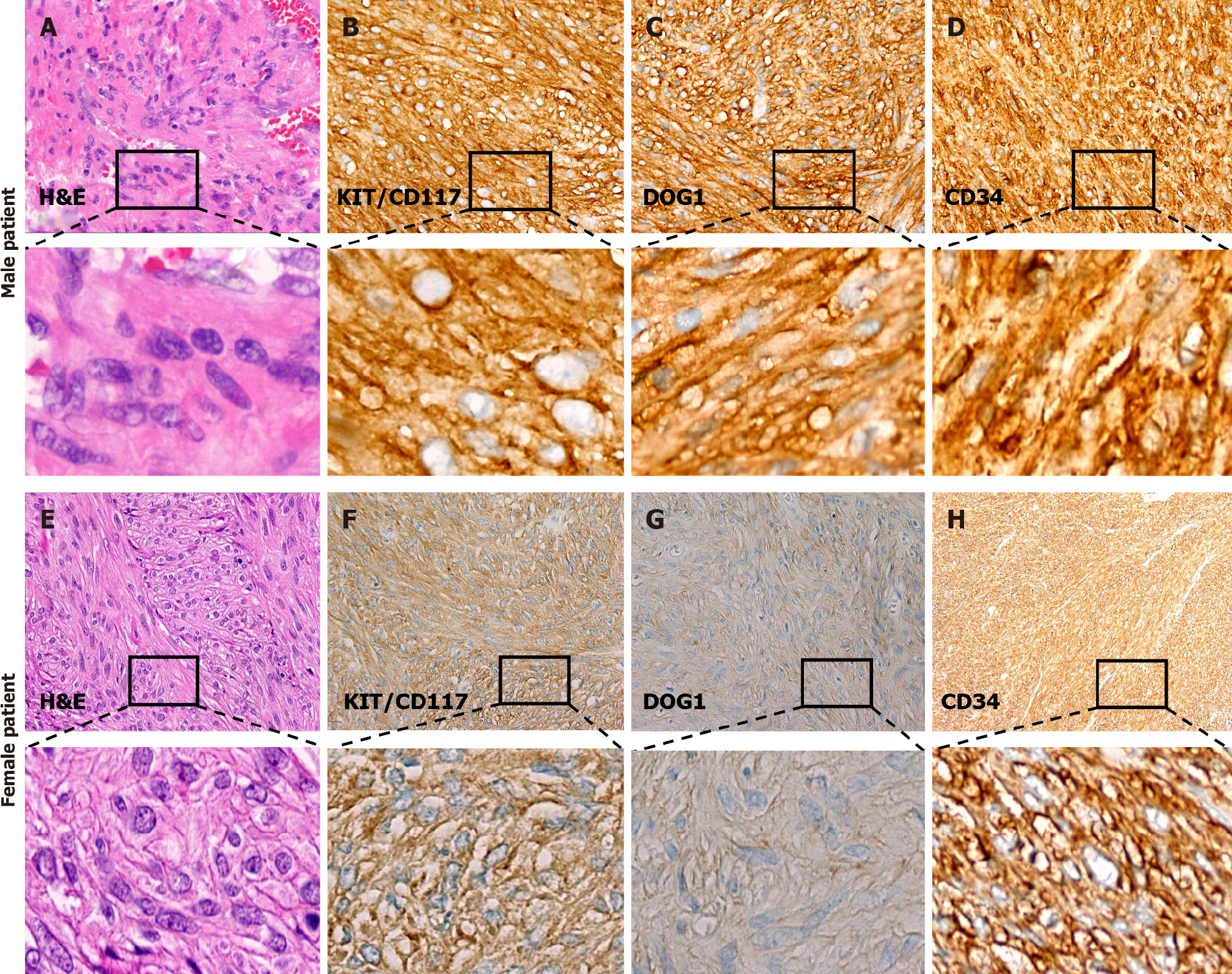
Figure 4 Histopathological and immunohistochemical examination of the lesions.
A: The lesions were stained with hematoxylin and eosin (H&E), revealing spindle-shaped cells arranged in bundles; B: KIT/CD117 showed positive immune responses; C: DOG1 showed positive immune responses; D: CD34 showed positive immune responses; E: The lesions were stained with H&E, revealing spindle-shaped cells arranged in bundles; F: KIT/CD117 showed positive immune responses; G: DOG1 showed positive immune responses; H: CD34 showed positive immune responses (original magnification, × 40).
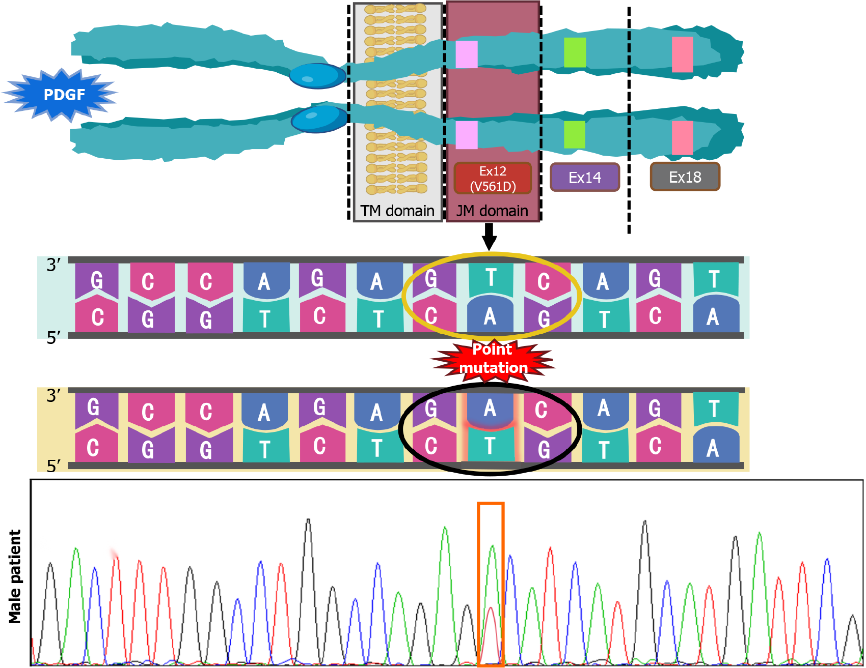
Figure 5 The results of gene detection in the male patient.
The excised lesion showed the presence of V561D mutation in exon 12 of the PDGFRA gene. The GTC to GAC conversion occurred at codon 561. TM: Transmembrane domain; JM: Intracellular juxtamembrane domain; PDGF: Platelet-derived growth factor.
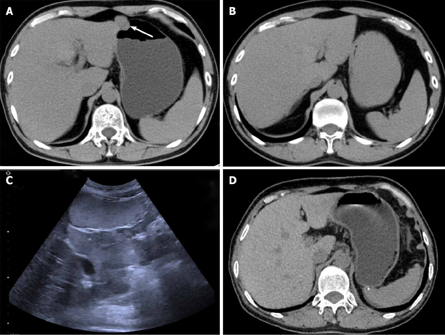
Figure 6 Postoperative follow-up examination results in September 2023.
Comparison of pre- and postoperative computed tomography (CT) images of the male patient. A: Preoperative CT plain scan of the whole abdomen with localized nodular shadows in the antrum of the stomach; B: Postoperative CT plain scan of the whole abdomen with postoperative changes in the distal stomach and no abnormal signs in the anastomosis; C: Gastrointestinal ultrasonography showing postoperative gastrointestinal stromal tumor, with no space-occupying lesions inside or outside the gastric lumen; D: Postoperative CT of the female patient showing postoperative changes after partial resection of the gastric fundus with no abnormal signs in the anastomosis.
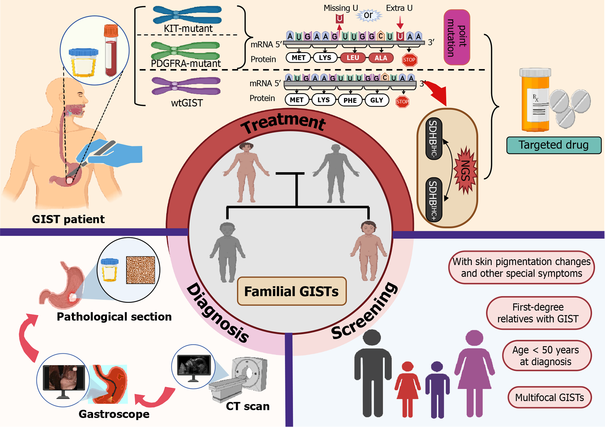
Figure 7 Familial gastrointestinal stromal tumors showing the diagnosis, treatment, and screening process.
GIST: Gastrointestinal stromal tumors; wtGISTs: Wild-type gastrointestinal stromal tumors; CT: Computed tomography; NGS: Next-generation sequencing; SDHB: Succinate dehydrogenase complex subunit; IHC: Immunohistochemical staining.
- Citation: Wang XK, Shen LF, Yang X, Su H, Wu T, Tao PX, Lv HY, Yao TH, Yi L, Gu YH. Two different mutational types of familial gastrointestinal stromal tumors: Two case reports. World J Gastrointest Oncol 2024; 16(9): 4028-4036
- URL: https://www.wjgnet.com/1948-5204/full/v16/i9/4028.htm
- DOI: https://dx.doi.org/10.4251/wjgo.v16.i9.4028


