Copyright
©The Author(s) 2024.
World J Gastrointest Oncol. Sep 15, 2024; 16(9): 3980-3993
Published online Sep 15, 2024. doi: 10.4251/wjgo.v16.i9.3980
Published online Sep 15, 2024. doi: 10.4251/wjgo.v16.i9.3980
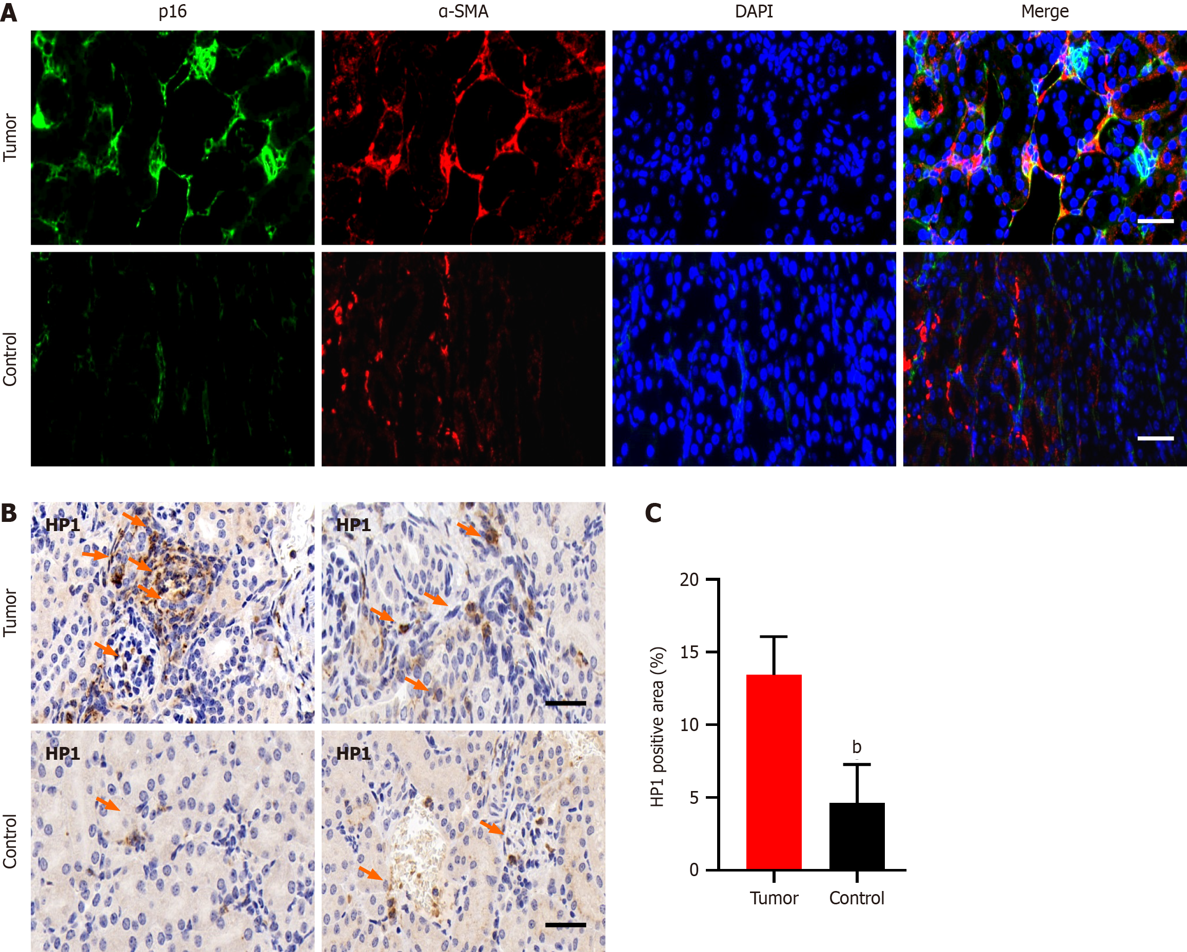
Figure 1 Senescent tumor-associated fibroblasts are present in pancreatic tumor tissue.
A: p16 was also highly expressed in regions with high α-SMA expression in pancreatic cancer tumor tissue; B and C: Pancreatic tumor tissues showed strong HP1 expression. The scale bar represents 20 µm. bP < 0.01.
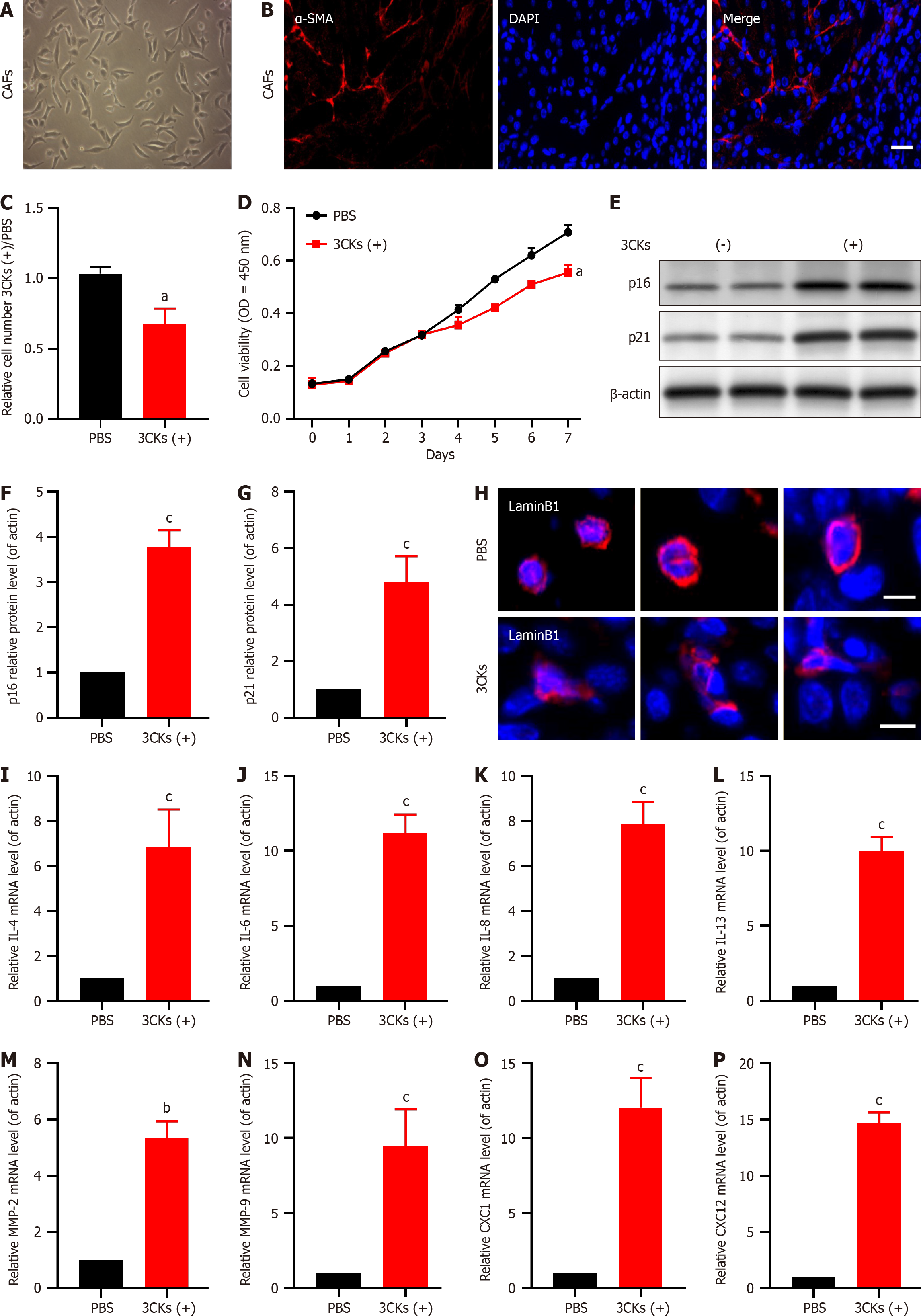
Figure 2 Stimulation with 3CKs induces senescence in cancer-associated fibroblasts.
A: Primary cultures of cancer-associated fibroblasts (CAFs) isolated from pancreatic tumor tissue were established; B: The isolated and cultured primary cells showed strong expression of α-SMA; C: Stimulation with 3CKs inhibited the growth of CAFs; D: The growth rate of CAFs significantly decreased after stimulation with 3CKs; E-G: The expression of p21 and p16 significantly increased after stimulating CAFs with 3CKs; H: The depletion of Lamin B1 was observed in 3CKs-treated CAFs; I-P: Treatment with 3CKs induced upregulation of senescence-associated secretory phenotype factors. The scale bar represents 20 µm. aP < 0.05, bP < 0.01, cP < 0.001.
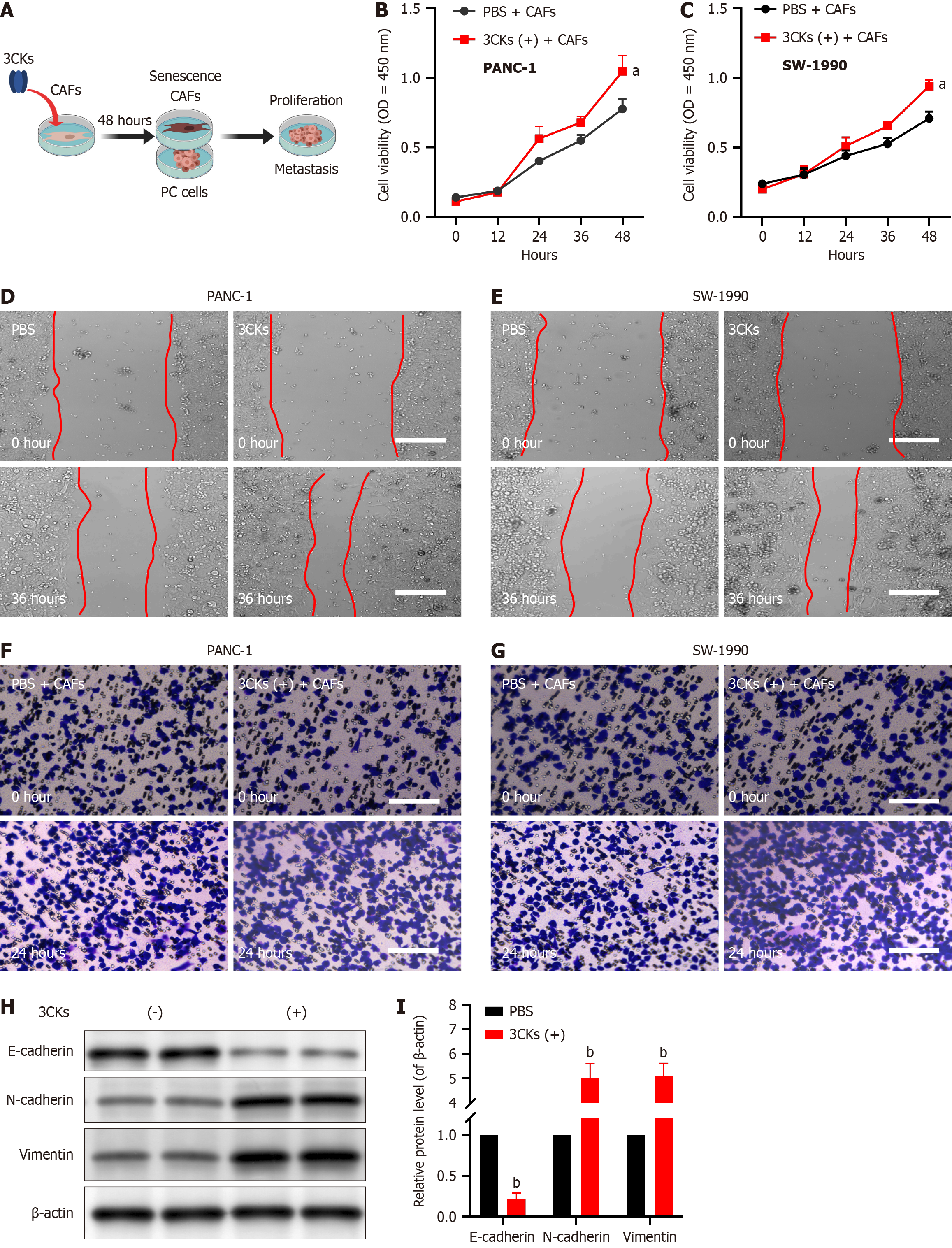
Figure 3 Senescent cancer-associated fibroblasts promote the proliferation and metastasis of pancreatic cancer cells.
A: Cancer-associated fibroblasts (CAFs) were cocultured with pancreatic cancer cells; B: The proliferation capacity of PANC1 cells was significantly enhanced when co-cultured with senescent CAFs; C: When co-cultured with senescent CAFs, the proliferative capacity of SW-1990 cells was significantly enhanced; D: The migration of PANC1 cells from the scratch edge to the center was more prominent when co-cultured with CAFs; E: SW-1990 cells exhibited enhanced migration towards the center of the scratch when cocultured with CAFs; F: PANC1 cells cocultured with CAFs had a greater invasion ability; G: SW-1990 cells cocultured with CAFs had a more extraordinary invasion ability; H and I: Epithelial-mesenchymal transition was induced in pancreatic cancer cells by senescent CAFs. The scale bar represents 20 µm. aP < 0.05, bP < 0.01, cP < 0.001. CAF: Cancer-associated fibroblasts; PBS: Phosphate-buffered saline.
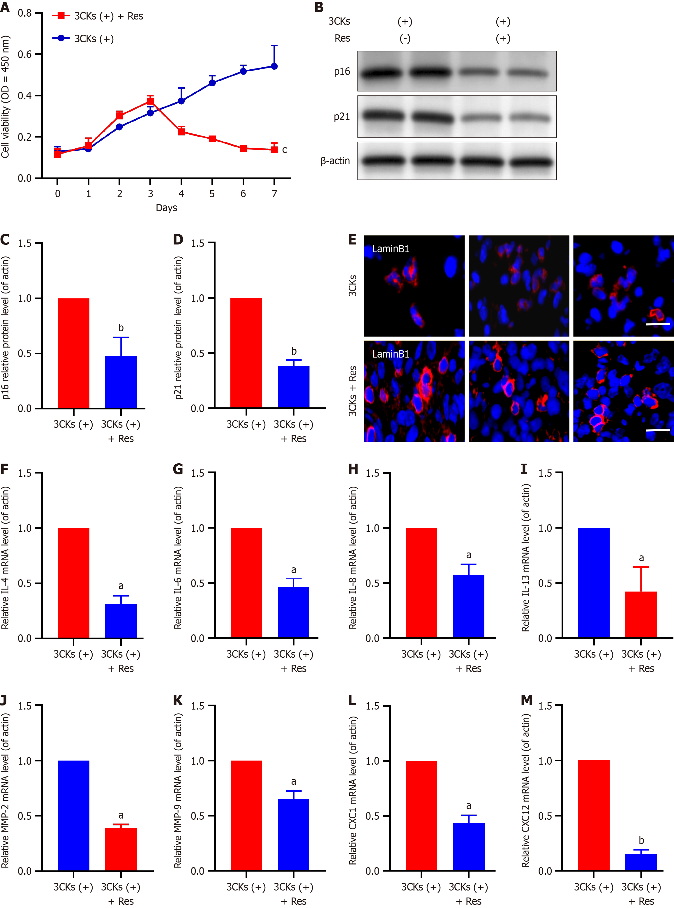
Figure 4 Resveratrol inhibits senescence in cancer-associated fibroblasts.
A: Resveratrol treatment resulted in the elimination of senescent cancer-associated fibroblasts; B-D: The expression of p21 and p16 was suppressed by resveratrol; E: Resveratrol intervention led to the restoration of Lamin B1 expression; F-M: There was a significant decrease in the expression of senescence-associated secretory phenotype factors. The scale bar represents 20 µm. aP < 0.05, bP < 0.01, cP < 0.001.
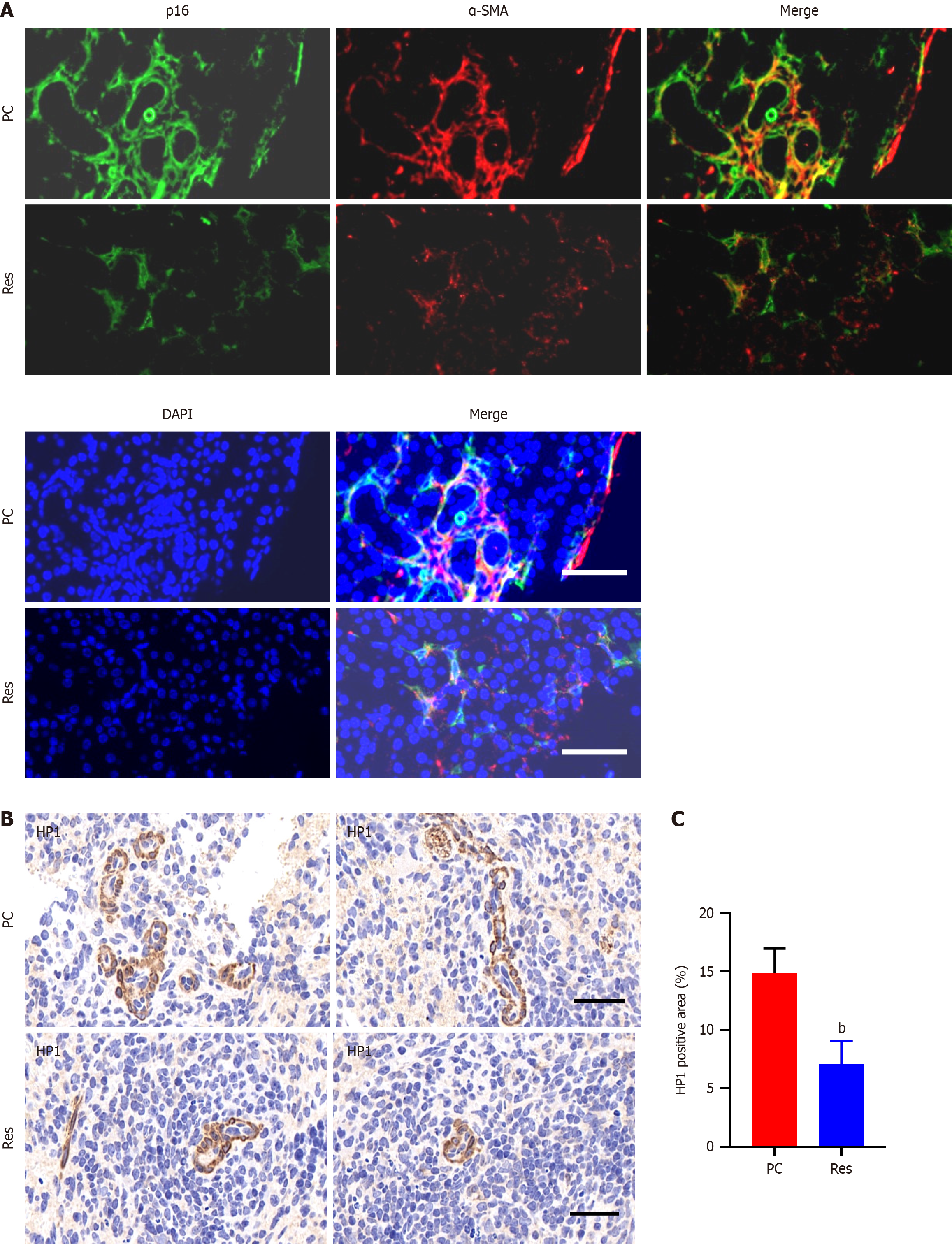
Figure 5 Resveratrol inhibits the expression of senescence markers.
A: The expression of p16 and α-SMA was suppressed by resveratrol treatment; B and C: The expression of HP-1 was inhibited by resveratrol. The scale bar represents 20 µm. bP < 0.01.
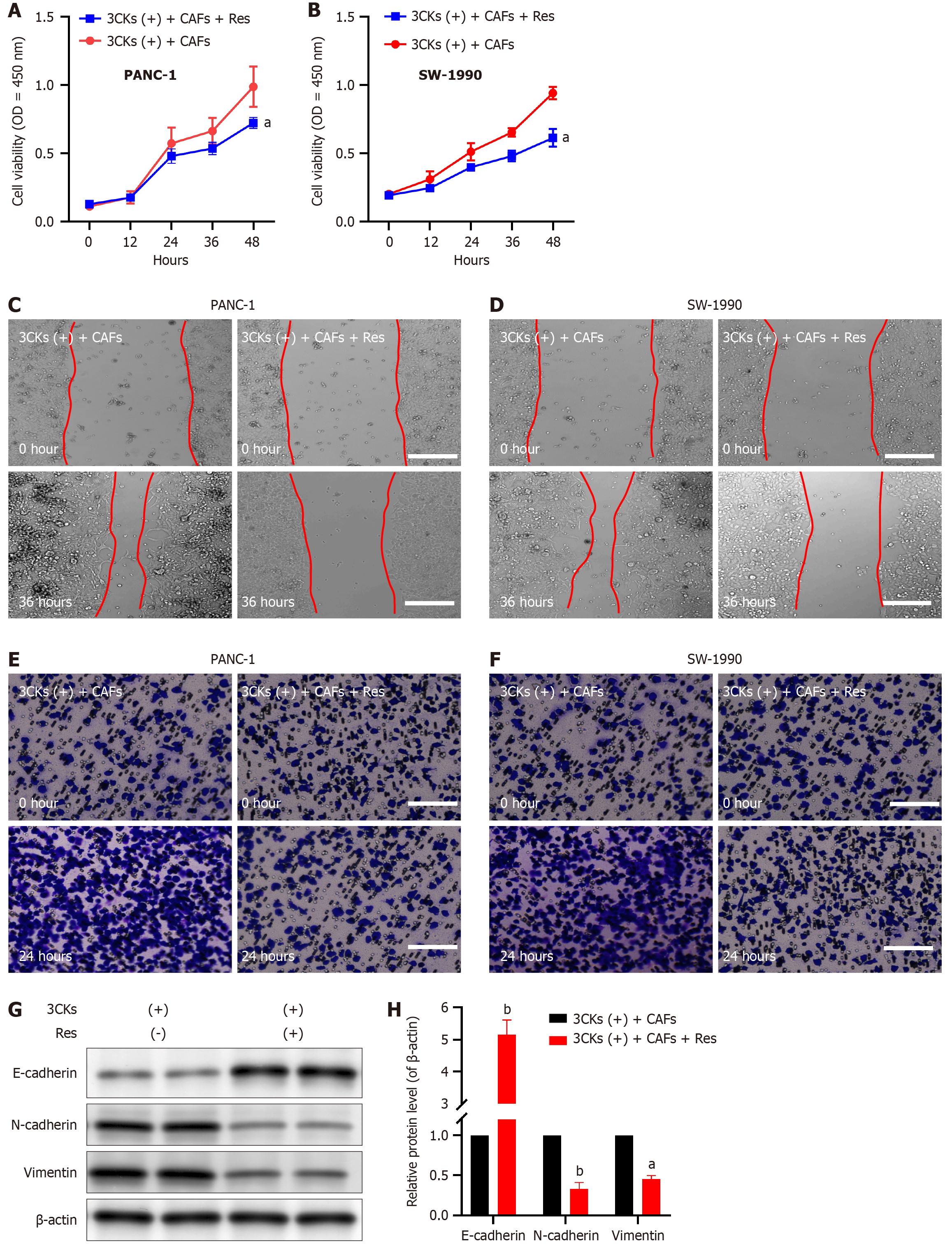
Figure 6 Resveratrol inhibits pancreatic cancer cell proliferation and metastasis.
A: Resveratrol effectively suppressed the proliferation of PANC-1 cells by inhibiting senescence in cancer-associated fibroblasts (CAFs); B: Resveratrol suppressed SW-1990 cell proliferation by inhibiting CAF senescence; C: The migratory ability of PANC-1 cells was inhibited by resveratrol; D: Resveratrol inhibited the migration of SW-1990 cells; E: Resveratrol inhibited the invasion of PANC-1 cells; F: Resveratrol inhibited the invasion of SW-1990 cells; G and H: After resveratrol intervention, there was evidence of epithelial-mesenchymal transition induction in pancreatic cancer cells. The scale bar represents 20 µm. aP < 0.05, bP < 0.01.
- Citation: Jiang H, Wang GT, Wang Z, Ma QY, Ma ZH. Resveratrol inhibits pancreatic cancer proliferation and metastasis by depleting senescent tumor-associated fibroblasts. World J Gastrointest Oncol 2024; 16(9): 3980-3993
- URL: https://www.wjgnet.com/1948-5204/full/v16/i9/3980.htm
- DOI: https://dx.doi.org/10.4251/wjgo.v16.i9.3980


