Copyright
©The Author(s) 2024.
World J Gastrointest Oncol. Aug 15, 2024; 16(8): 3723-3731
Published online Aug 15, 2024. doi: 10.4251/wjgo.v16.i8.3723
Published online Aug 15, 2024. doi: 10.4251/wjgo.v16.i8.3723
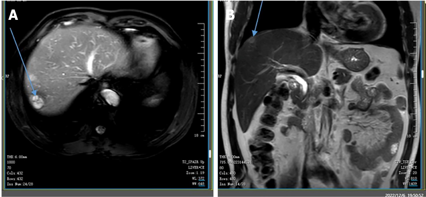
Figure 1 Liver magnetic resonance images.
Magnetic resonance images performed at Yichang Central People's Hospital on February 9, 2022. Well-circumscribed, heterogeneous, and hypodense masses are located in the right posterior lobe of the liver, measuring 2.1 cm × 2.0 cm (indicated by the arrow). A: Transverse view; B: Coronal view.
Figure 2 Positron emission tomography-computed tomography images from March 2022 showing multiple masses in the mesentery, omentum, and peritoneum with increased abnormal tracer metabolism.
Figure 3 Pathology after laparoscopic exploration of the greater omentum.
The tumor cells are spindle-shaped or cluster-shaped, with oval and deeply stained nuclei. A: Shows hematoxylin and eosin staining under a × 200 microscope; B-D: Show immunohistochemical staining: Cytoplasmic vacuoles are observed in some cells. Tumor cell membranes are positive for Caveolin-1, and the nuclei are positive for GATA3 and Ki-67 (Li > 70%).
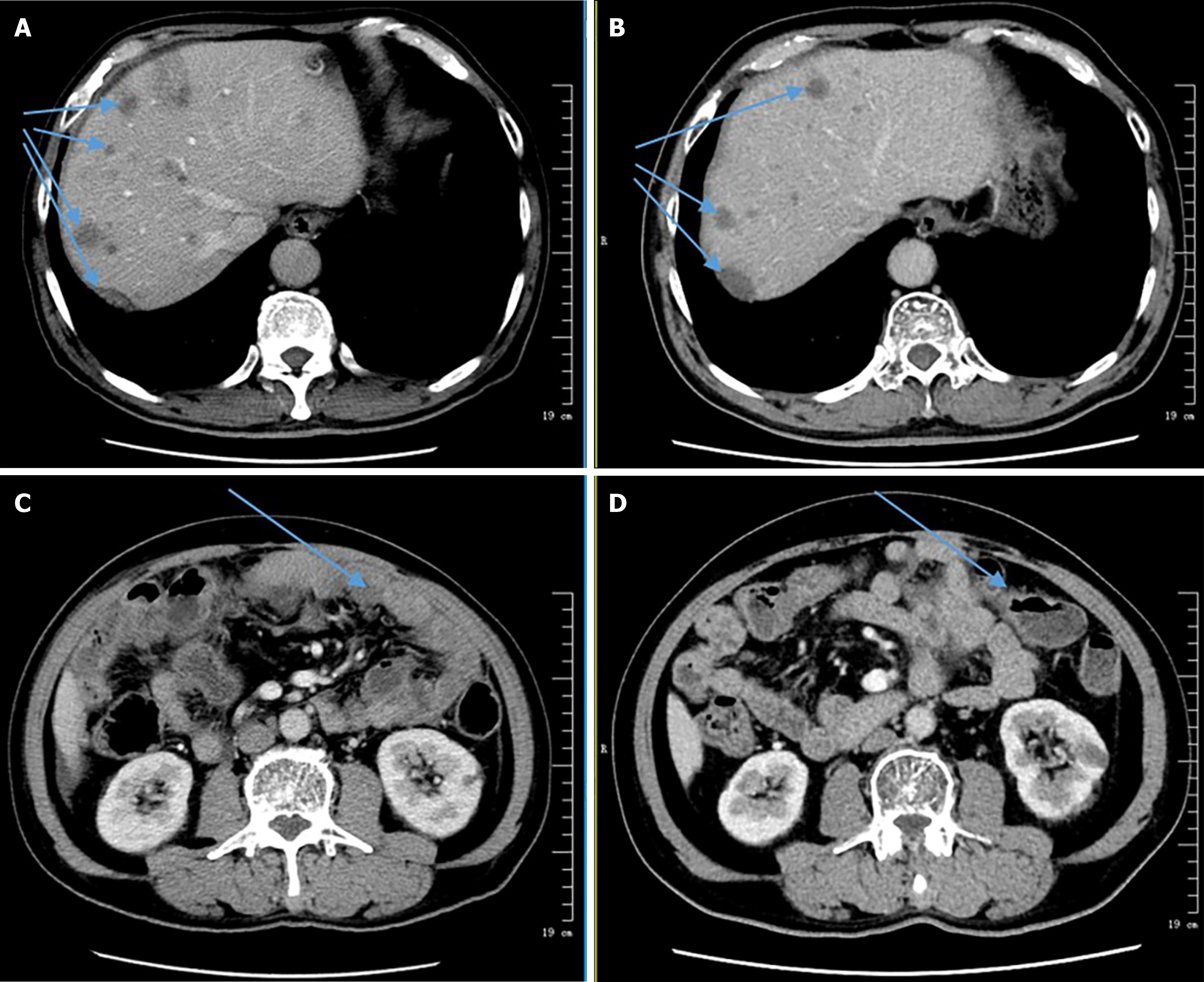
Figure 4 Comparison of Computed tomography.
A: Computed tomography (CT) images from May 18, 2022; B: CT images from September 14, 2022; A and B: The total number of liver lesions has decreased, and some lesions have reduced in enhancement compared to the previous images (indicated by the arrow); C: CT images from May 18, 2022; D: CT images from September 14, 2022; C and D: Showing decreased peritoneal thickening, reduced omentum opacity, and decreased size of some nodules in the abdominal cavity. There is also a reduction in abdominal and pelvic fluid accumulation (indicated by the arrow).
- Citation: Feng Q, Yu W, Feng JH, Huang Q, Xiao GX. Jejunal sarcomatoid carcinoma: A case report and review of literature. World J Gastrointest Oncol 2024; 16(8): 3723-3731
- URL: https://www.wjgnet.com/1948-5204/full/v16/i8/3723.htm
- DOI: https://dx.doi.org/10.4251/wjgo.v16.i8.3723


