Copyright
©The Author(s) 2024.
World J Gastrointest Oncol. Jul 15, 2024; 16(7): 3331-3340
Published online Jul 15, 2024. doi: 10.4251/wjgo.v16.i7.3331
Published online Jul 15, 2024. doi: 10.4251/wjgo.v16.i7.3331
Figure 1 Hematoxylin and eosin staining of primary gastric cancer.
This specimen has lost its normal gastric architecture, with multiple tumor cells presenting as solid bands of varying sizes. A: Original magnification × 200; B: Original magnification × 100.
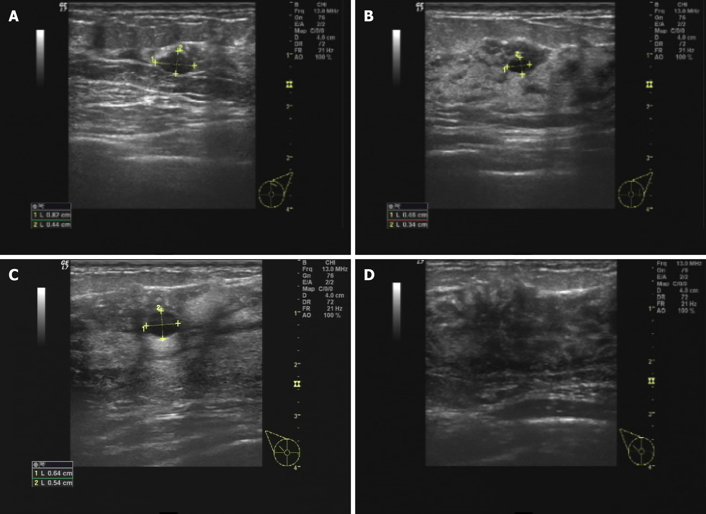
Figure 2 Ultrasound images.
A: A nodule in the left breast at 1 o’clock, measuring approximately 8 mm × 4 mm; B: A nodule in the left breast at 3 o’clock, measuring approximately 5 mm × 3 mm; C: A nodule in the right breast at 10 o’clock, measuring approximately 6 mm × 5 mm; D: Multiple calcified foci in the upper outer quadrant of the right breast.
Figure 3 Bilateral breast mammogram.
The expanded box shows the calcification point. The density of both breasts is heterogeneous, with scattered lamellar, flocculent and granular high-density shadows and multiple punctate calcifications between the breasts, with a more scattered distribution in the left breast and a partially clustered distribution in the right breast, predominantly in the right upper quadrant. A and B: A mammogram of the left breast; C and D: A mammogram of the right breast.
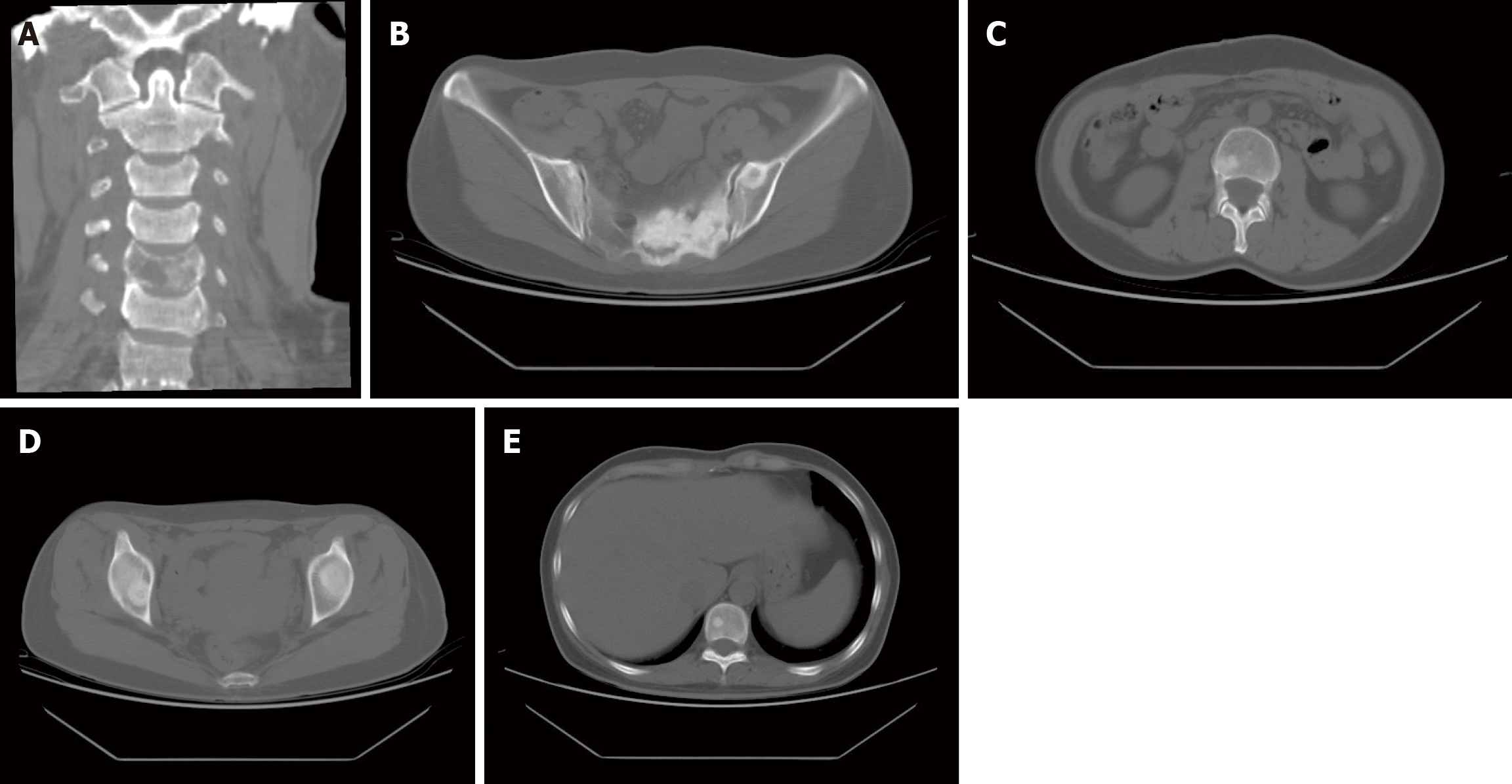
Figure 4 Computed tomography imaging images.
A: Bony destruction of the C5 vertebrae, considering tumour metastasis; B: Metastasis in the left ilium; C: Metastasis in the lumbar spine; D: Metastasis in the thoracic spine; E: Metastasis in the right acetabulum.
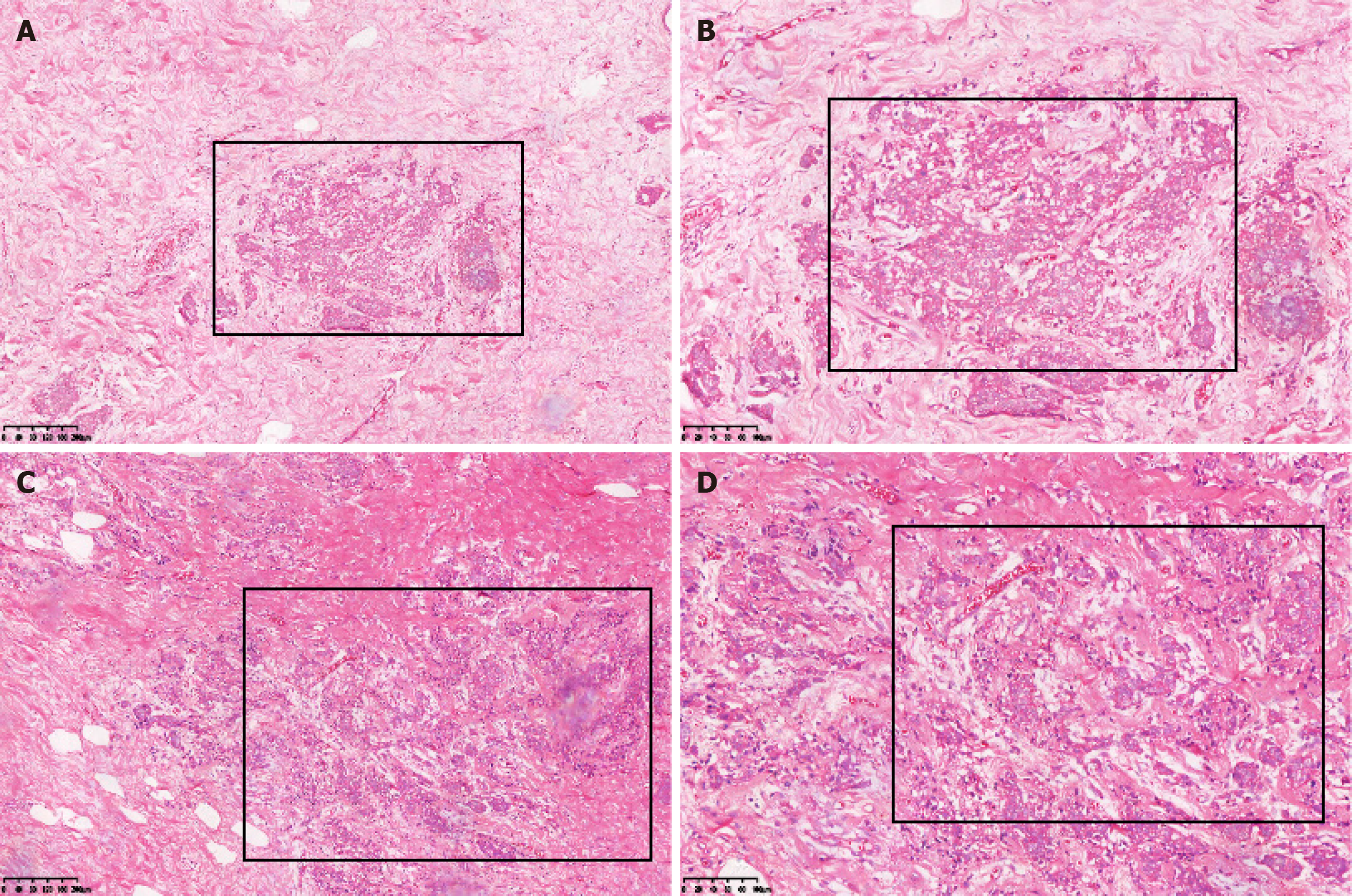
Figure 5 Hematoxylin and eosin staining of metastatic breast cancer.
There were diffuse infiltrating epithelioid cells filled in the interstitium of the mammary gland. A: Left breast (original magnification × 100); B: Left breast (original magnification × 200); C: Right breast (original magnification × 100); D: Right breast (original magnification × 200).
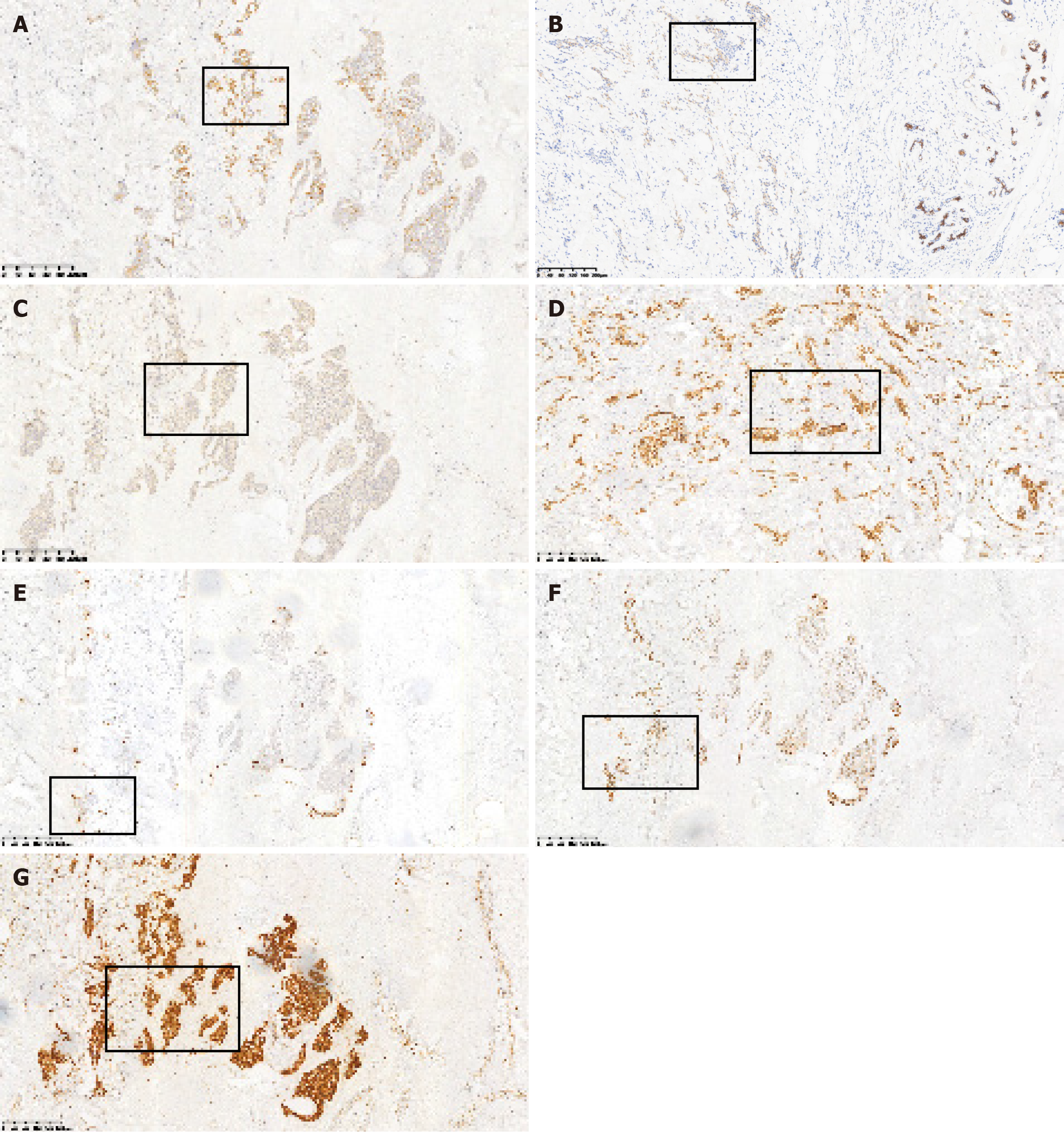
Figure 6 Positive immunohistochemical staining indicators.
A: 34E12; B: Cytokeratin (CK) 7; C: CK56; D: E-cad; E: Ki67; F: Tumor protein 53; G: P120.
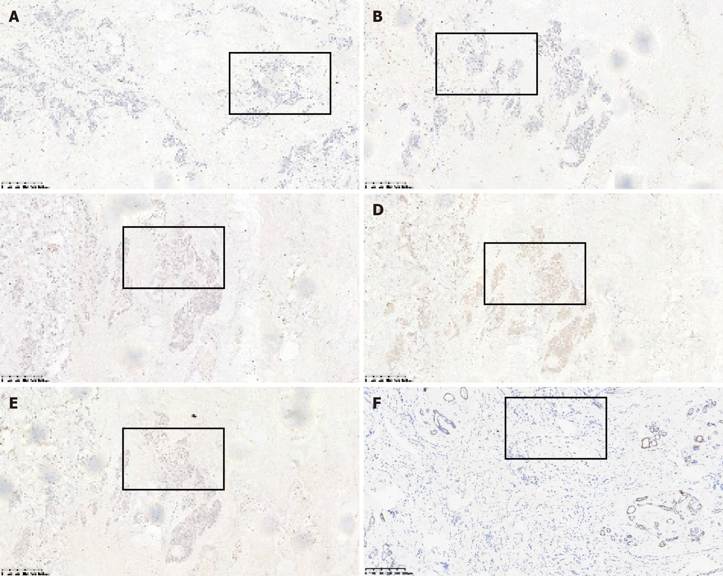
Figure 7 Negative immunohistochemical staining indicators.
A: Cytokeratin 20; B: Epidermal growth factor receptor (EGFR); C: Estrogen receptor; D: Human EGFR 2; E: Progesterone receptor; F: GATA binding protein 3.
- Citation: Liu JH, Dhamija G, Jiang Y, He D, Zhou XC. Gastric cancer metastatic to the breast: A case report. World J Gastrointest Oncol 2024; 16(7): 3331-3340
- URL: https://www.wjgnet.com/1948-5204/full/v16/i7/3331.htm
- DOI: https://dx.doi.org/10.4251/wjgo.v16.i7.3331


