Copyright
©The Author(s) 2024.
World J Gastrointest Oncol. Jun 15, 2024; 16(6): 2531-2540
Published online Jun 15, 2024. doi: 10.4251/wjgo.v16.i6.2531
Published online Jun 15, 2024. doi: 10.4251/wjgo.v16.i6.2531
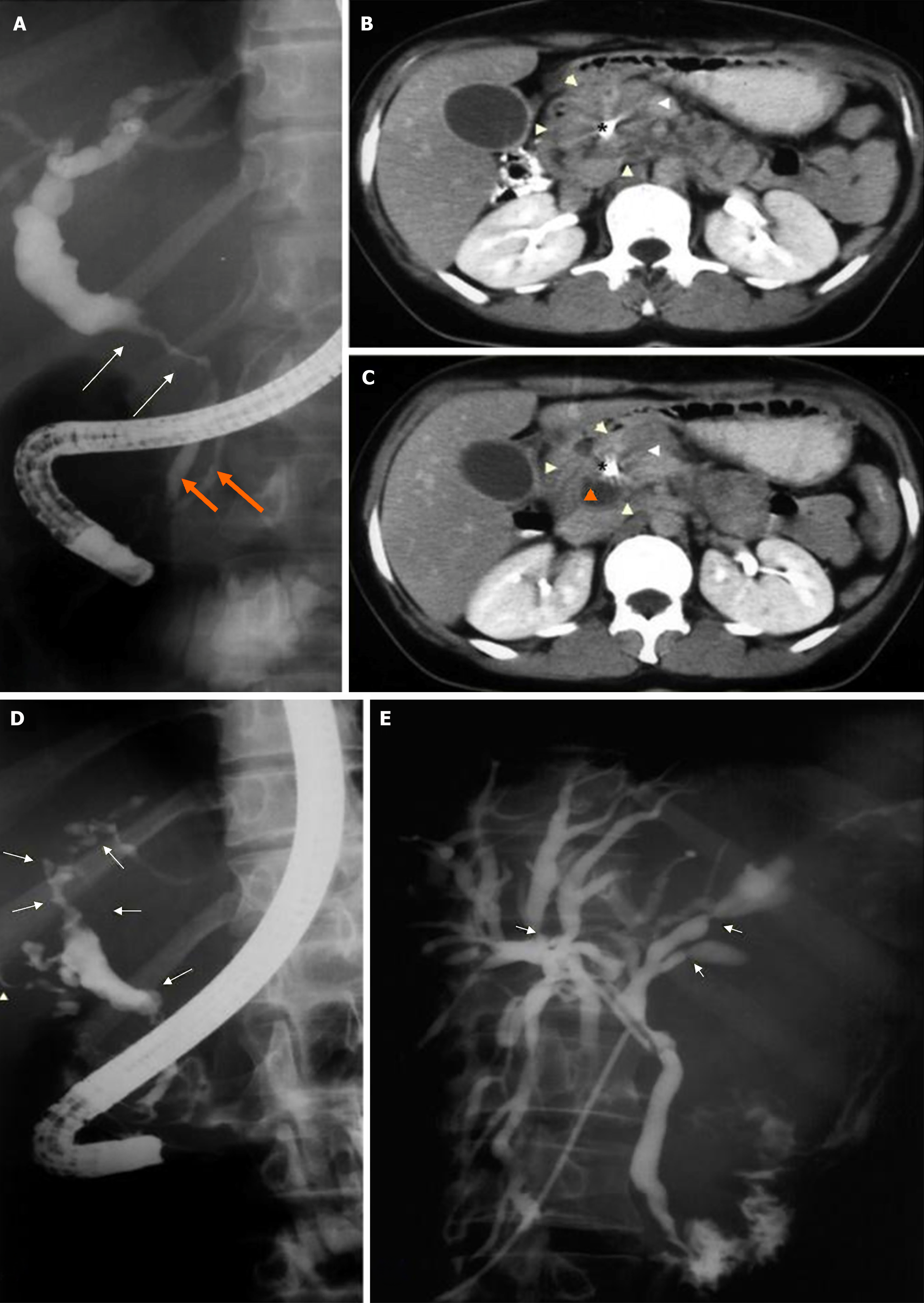
Figure 1 Paracoccidioidomycosis-related extrahepatic cholestasis mimicking cholangiocarcinoma and primary sclerosing cholangitis.
A: Endoscopic retrograde cholangiopancreatography showing abrupt stenosis of the common bile duct and irregular walls (white arrows). The intrahepatic bile duct is dilated, while the distal common bile duct and pancreatic duct have normal calibers (orange arrows); B and C: Computed tomography after placement of a plastic stent in the bile duct (asterisk*) showing enlargement of the pancreas head (white arrowheads) and a cystic component (orange arrow); D: Endoscopic retrograde cholangiopancreatography before treatment showing irregularities in the wall of the intra and extrahepatic bile ducts, with areas of segmental stenosis and dilation (white arrows), similar to primary sclerosing cholangitis; E: T-tube cholangiography 1 month after drug treatment showing improvement of wall irregularities and areas of stenosis. There is slight residual dilation and restricted areas of stenosis (white arrows).
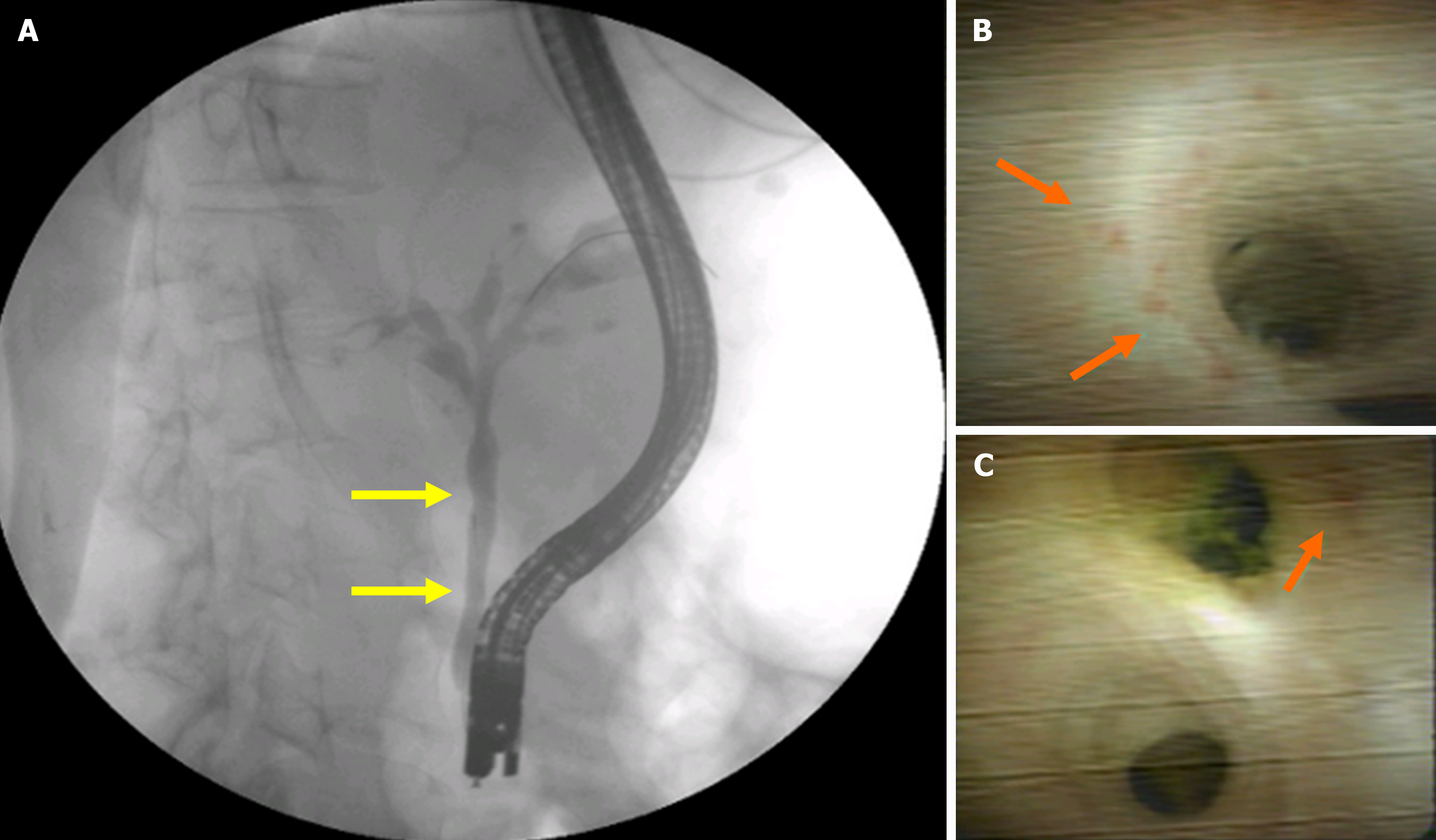
Figure 2 Cholestasis, consumptive syndrome, and jaundice associated with blastomycosis.
A: Endoscopic retrograde cholangiopancreatography revealing irregularities in the wall of the common distal bile duct (yellow arrows); B and C: Images obtained by cholangioscopy biopsy of the distal common bile duct wall, which shows some superficial irregularity (orange arrows).
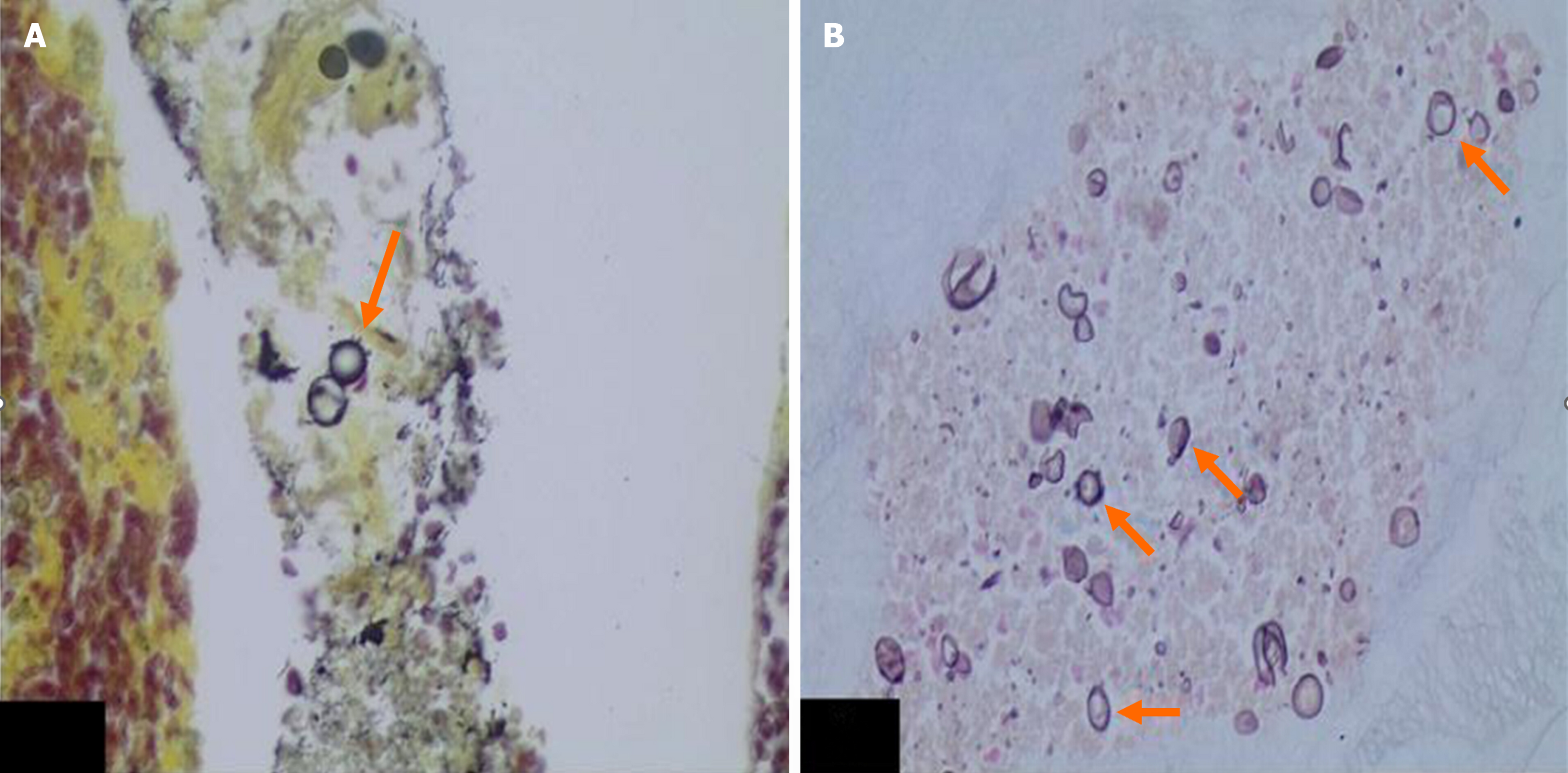
Figure 3 Cholangioscopy biopsy.
A and B: Presence of rounded fungal structures, detected by the Gomori’s methenamine silver (GMS) staining method, with multiple sporulation (orange arrows) compatible with paracoccidioidomycosis (GMS, 400 ×).
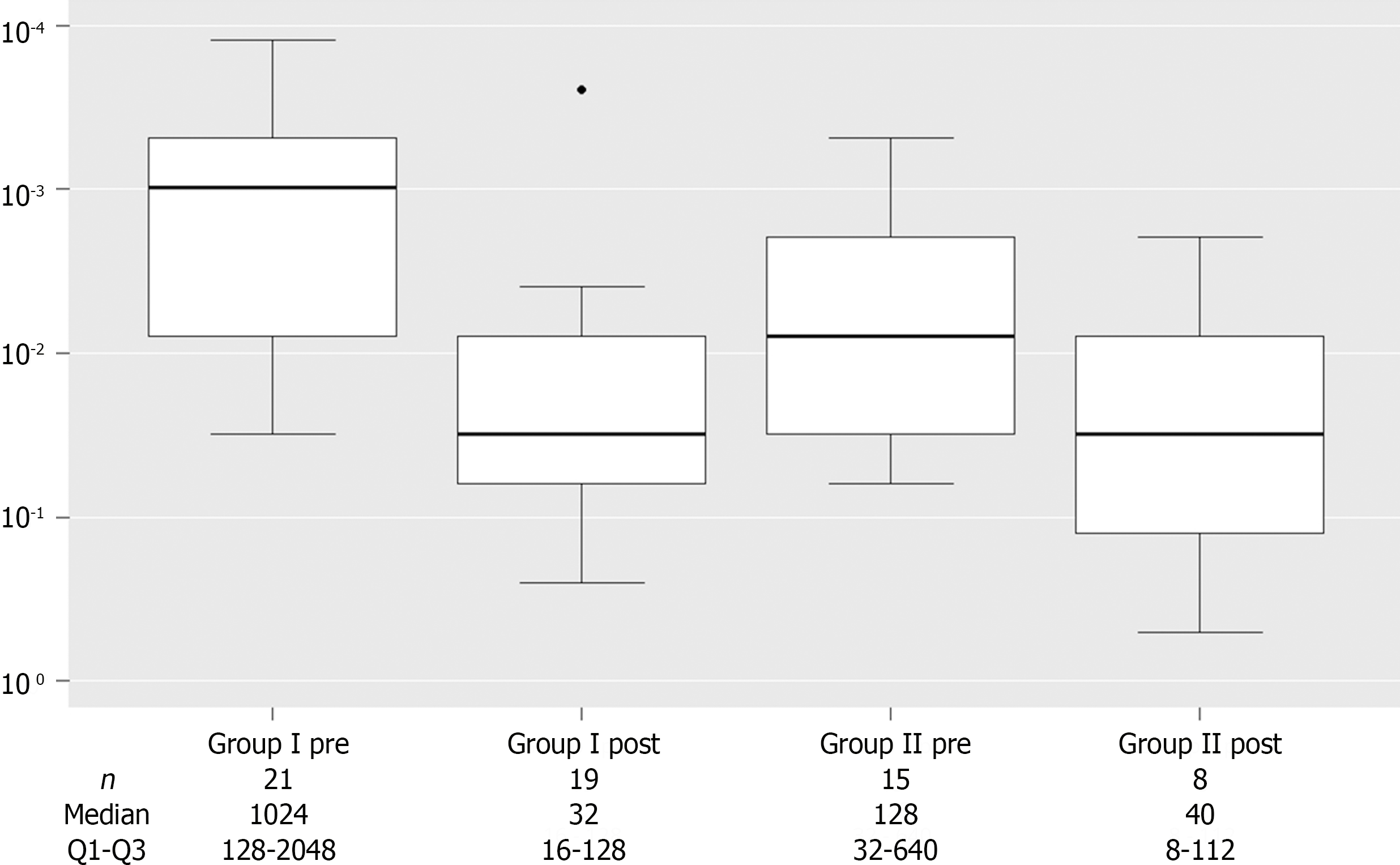
Figure 4
Pre- and post-treatment counterimmunoelectrophoresis values in patients with paracoccidioidomycosis and extrahepatic cholestasis allocated to groups 1 and 2.
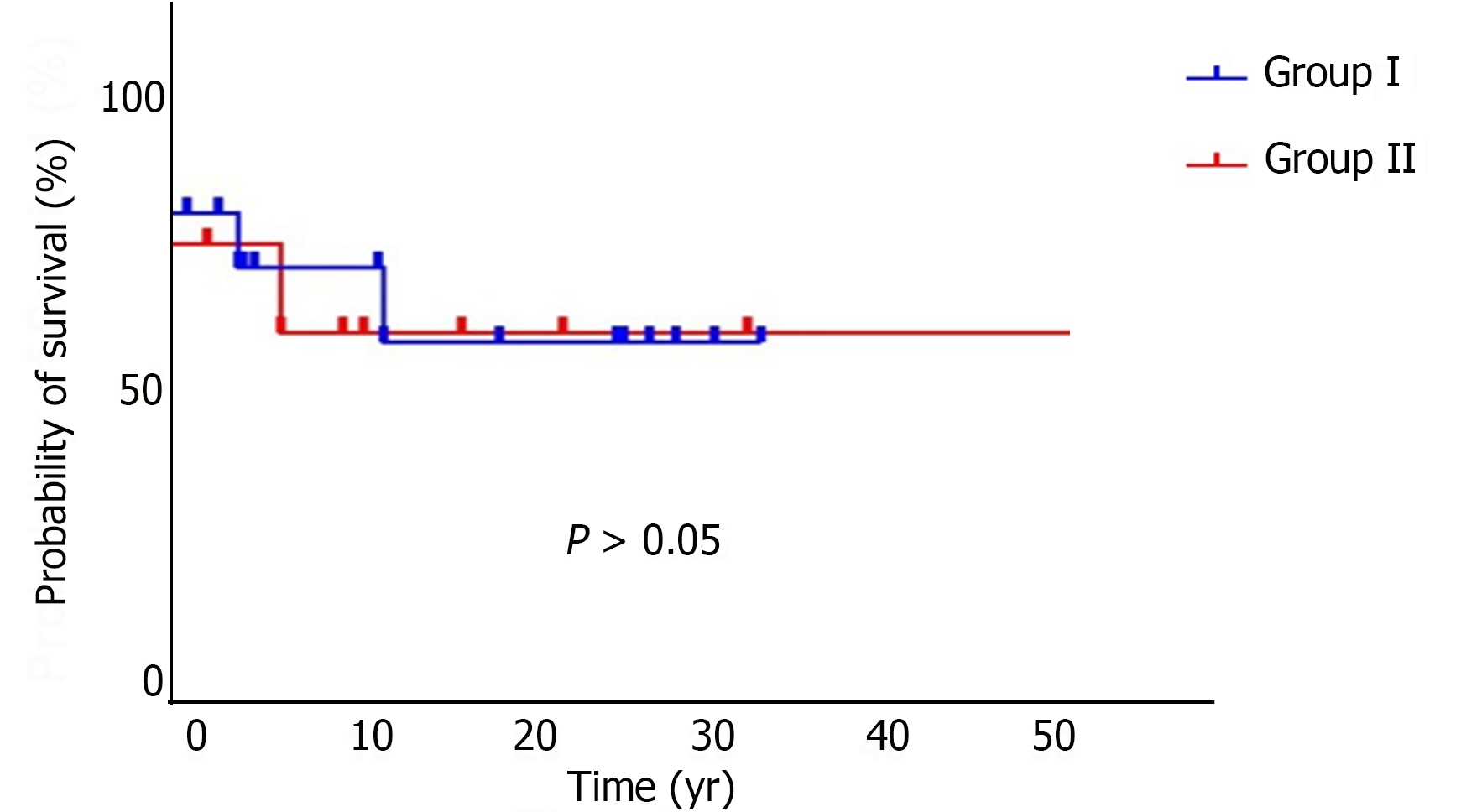
Figure 5
Survival estimates by group.
- Citation: dos Santos JS, de Moura Arrais V, Rosseto Ferreira WJ, Ribeiro Correa Filho R, Brunaldi MO, Kemp R, Sankanrakutty AK, Elias Junior J, Bellissimo-Rodrigues F, Martinez R, Zangiacomi Martinez E, Ardengh JC. Extrahepatic cholestasis associated with paracoccidioidomycosis: Challenges in the differential diagnosis of biliopancreatic neoplasia. World J Gastrointest Oncol 2024; 16(6): 2531-2540
- URL: https://www.wjgnet.com/1948-5204/full/v16/i6/2531.htm
- DOI: https://dx.doi.org/10.4251/wjgo.v16.i6.2531


