Copyright
©The Author(s) 2024.
World J Gastrointest Oncol. Jun 15, 2024; 16(6): 2476-2486
Published online Jun 15, 2024. doi: 10.4251/wjgo.v16.i6.2476
Published online Jun 15, 2024. doi: 10.4251/wjgo.v16.i6.2476
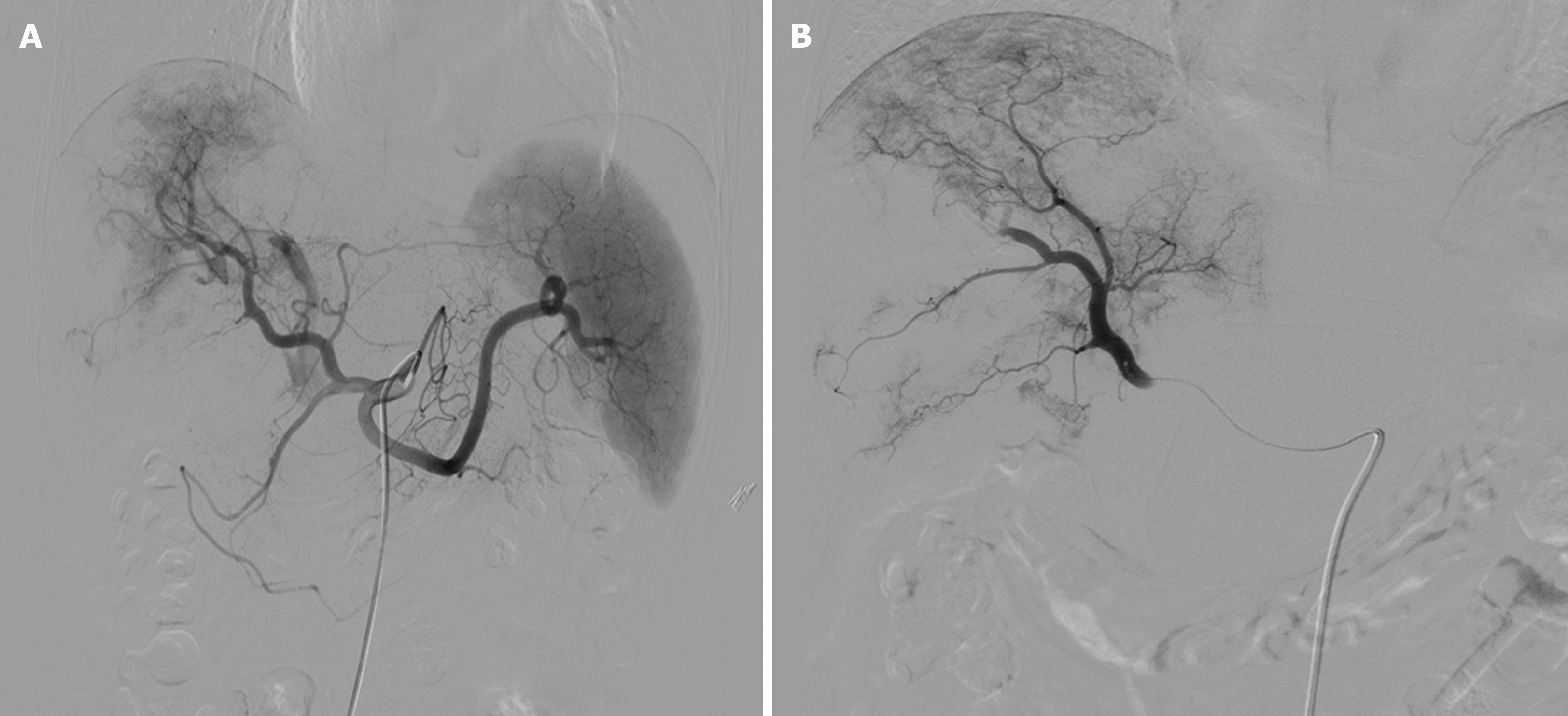
Figure 1 Tumor staining.
A: Abdominal arteriogram showing the arteries supplying the tumor, the tumor itself, portal vein tumor thrombus staining, and a portal fistula; B: Cessation of embolism with stasis in the artery supplying the tumor and the disappearance of tumor staining.
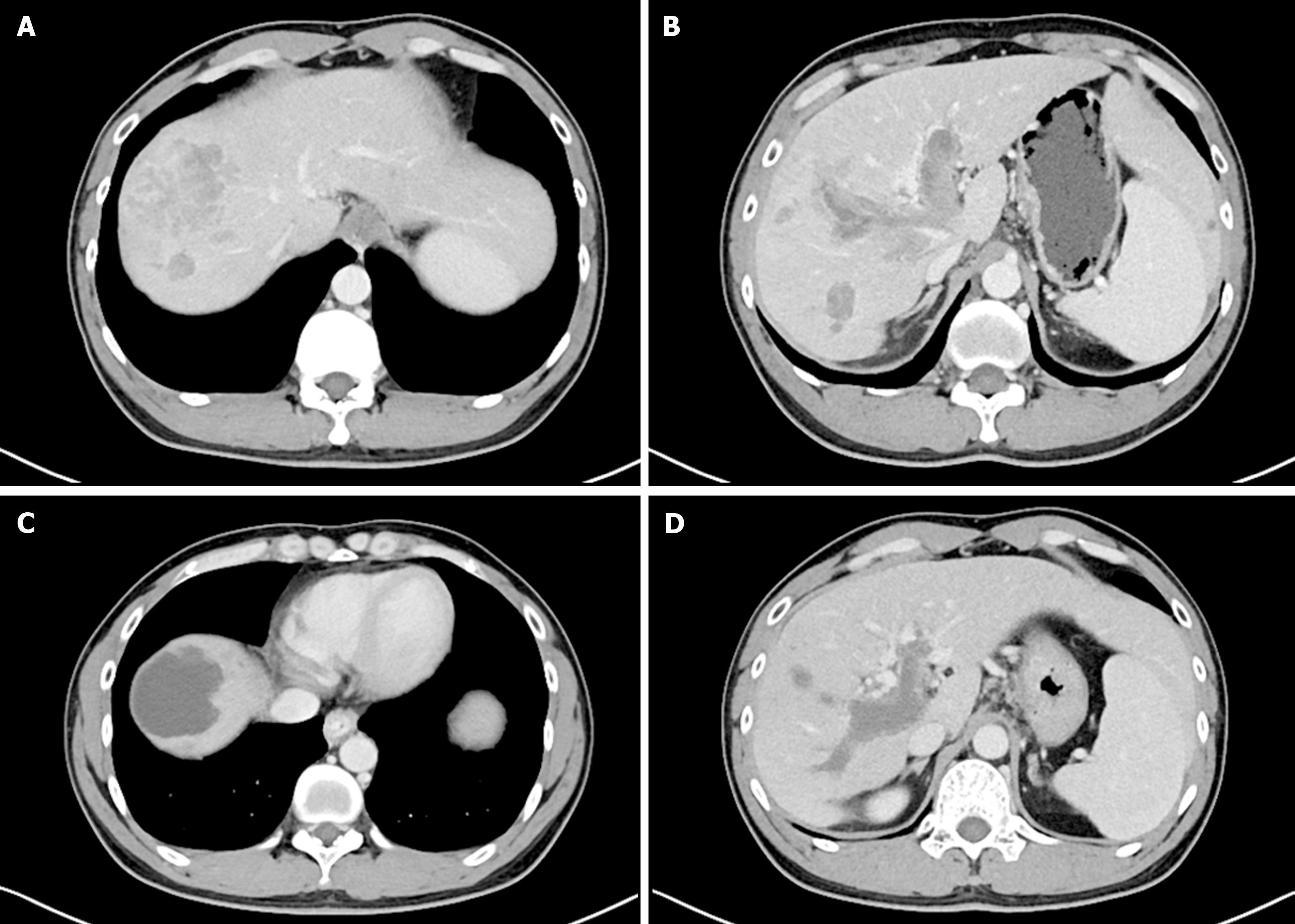
Figure 2 The patient was treated with drug-eluting bead transcatheter arterial chemoembolization for hepatocellular carcinoma with portal vein tumor thrombus.
A and B: Visible intrahepatic mass (A) and obvious enhancement of portal vein tumor thrombus (PVTT) III (B); C and D: After drug-eluting bead transcatheter arterial chemoembolization, the mass (C) and PVTT (D) showed extensive necrosis (C), and enhanced computed tomography showed a few enhanced lesions in the arterial phase: Modified response evaluation criteria in solid tumors efficacy was evaluated for partial response.
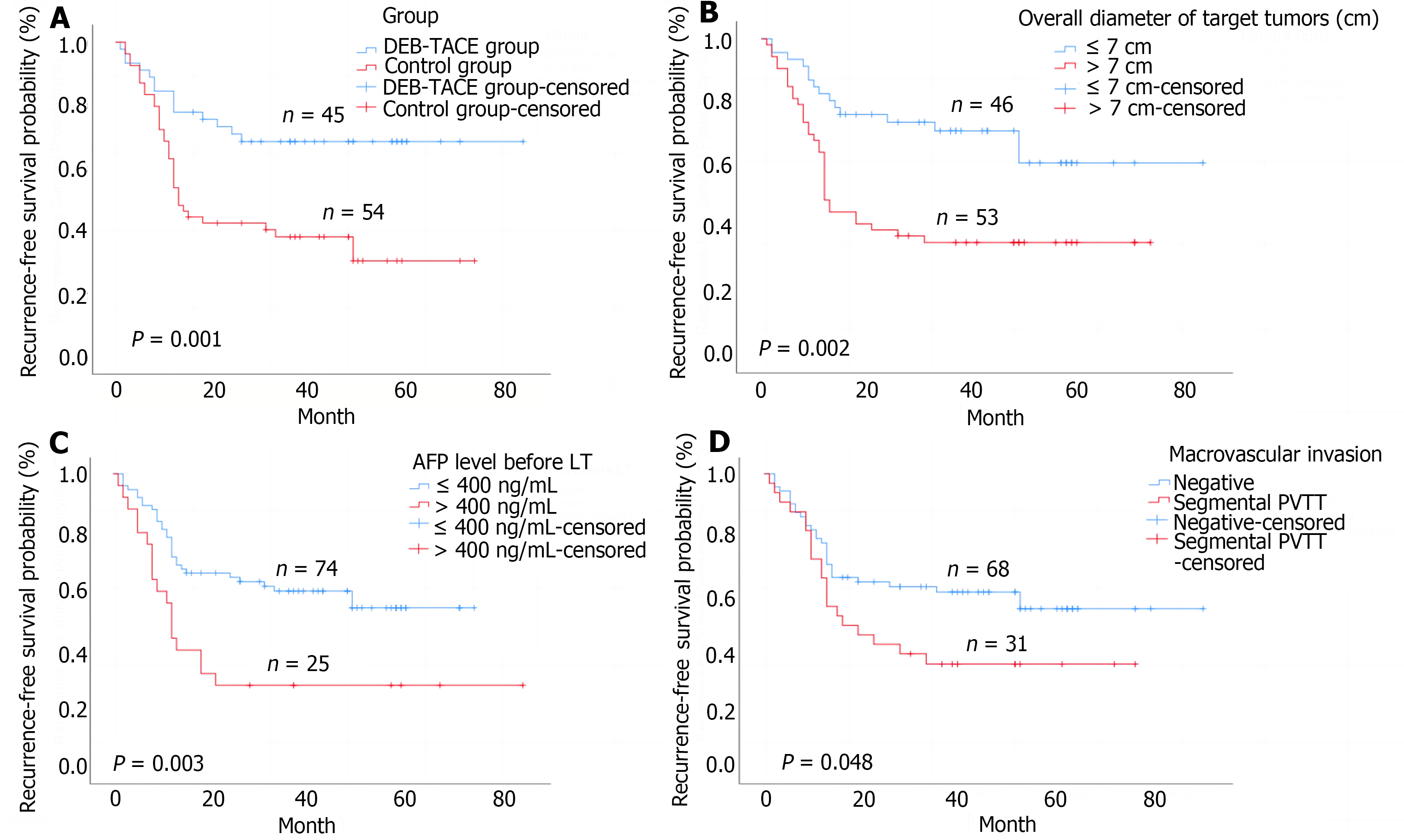
Figure 3 Kaplan-Meier analysis of survival outcomes in patients.
A: Recurrence-free survival comparisons; B: Overall diameter of target tumors; C: Alpha-fetoprotein level before liver transplantation; D: Macrovascular invasion. DEB-TACE: Drug-eluting bead transcatheter arterial chemoembolization; AFP: Alpha-fetoprotein; LT: Liver transplantation.
Figure 4 Kaplan-Meier analysis of survival outcomes in patients.
A: Overall survival comparisons; B: Overall diameter of target tumors. DEB-TACE: Drug-eluting bead transcatheter arterial chemoembolization.
Figure 5 Two-year recurrence-free survival rates and overall survival rates comparisons in subgroup analyses.
A: Patients without portal vein tumor thrombus (PVTT); B: Patients with segmental PVTT.
- Citation: Ye ZD, Zhuang L, Song MC, Yang Z, Zhang W, Zhang JF, Cao GH. Drug-eluting bead transarterial chemoembolization as neoadjuvant therapy pre-liver transplantation for advanced-stage hepatocellular carcinoma. World J Gastrointest Oncol 2024; 16(6): 2476-2486
- URL: https://www.wjgnet.com/1948-5204/full/v16/i6/2476.htm
- DOI: https://dx.doi.org/10.4251/wjgo.v16.i6.2476


