Copyright
©The Author(s) 2024.
World J Gastrointest Oncol. May 15, 2024; 16(5): 1821-1832
Published online May 15, 2024. doi: 10.4251/wjgo.v16.i5.1821
Published online May 15, 2024. doi: 10.4251/wjgo.v16.i5.1821
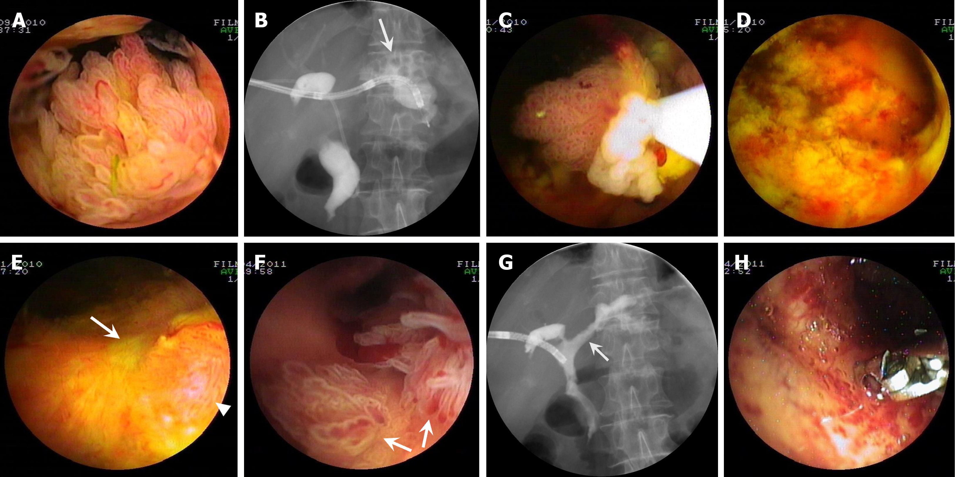
Figure 1 Percutaneous transhepatic cholangioscopy-assisted biliary polypectomy for mucin-hypersecreting cast-like type intraductal papillary neoplasm of the bile duct with therapeutic success in patient one.
A: Percutaneous transhepatic cholangioscopy (PTCS) showing frond-like tumors with vascular images in the hilar bile duct; B: Cholangiography showing multiple 1-cm sized stones in the left intrahepatic duct (arrow); C: PTCS showing a frond-like tumor that was being resected with the snare at the lower right in the visual field under PTCS; D: The cholangioscopic view of the location where the tumor was successfully resected with PTCS assisted biliary polypectomy (PTCS-BP); E: A scar (arrow) forming with a local minimal proliferated tumor (arrowhead) 2 wk after the last PTCS-BP; F: PTCS showing newly growing leaf-like tumors (arrows); G: Cholangiography showing a reduced biliary tract caliber, with a stenotic stiff and rigid ductal wall suspected of the existence of an invasive lesion in the left hepatic duct (arrow); H: Biopsies being taken from the site suspected of the existence of an invasive lesion 5 months after PTCS-BP.
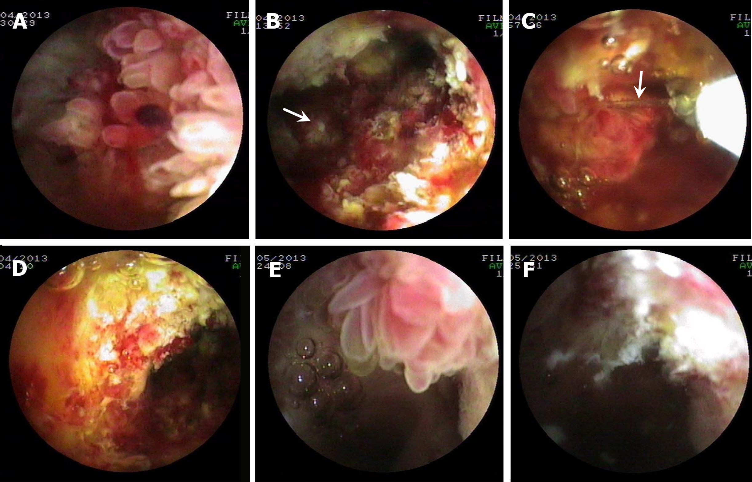
Figure 2 Percutaneous transhepatic cholangioscopy-assisted biliary polypectomy for recurrent mucin-hypersecreting cast-like type intraductal papillary neoplasm of the bile duct with therapeutic success in patient two who received photodynamic therapy for postoperative recurrent intraductal papillary neoplasm of the bile duct 11 months prior to Percutaneous transhepatic cholangioscopy-assisted biliary polypectomy.
A and B: Percutaneous transhepatic cholangioscopy (PTCS) showing the multiple papillary tumors and the locations (the arrow in Figure B refers to a resected piece of the tumor tissue) where the tumors were resected with PTCS-assisted biliary polypectomy (PTCS-BP), respectively; C and D: PTCS showing a polypoid tumor snared (the arrow in Figure C points to the snare steel wire) and the location where the tumor was resected with PTCS-BP, respectively; E and F: PTCS showing a frond-like projection and the locations where the tumors were resected with PTCS-BP, respectively; A, C, and E: PTCS showing the multiple papillary, a polypoid and frond-like tumors, respectively; B, D, and F: PTCS showing the locations where the tumor was successfully resected with PTCS-BP.
Figure 3 Percutaneous transhepatic cholangioscopy-assisted biliary polypectomy for non-mucin-producing polypoid type intraductal papillary neoplasm in the intrahepatic bile duct with therapeutic success in patient three.
A: Percutaneous transhepatic cholangioscopy (PTCS) showing a 1-cm sized polypoid mass with no obvious vascularity; B: A far view of the mass (from far view to observe the tumor); C: The tumor ready to be snared and resected under the direct visualization of PTCS (methylene blue staining); D: The location where the tumor was successfully resected with PTCS-assisted biliary polypectomy.
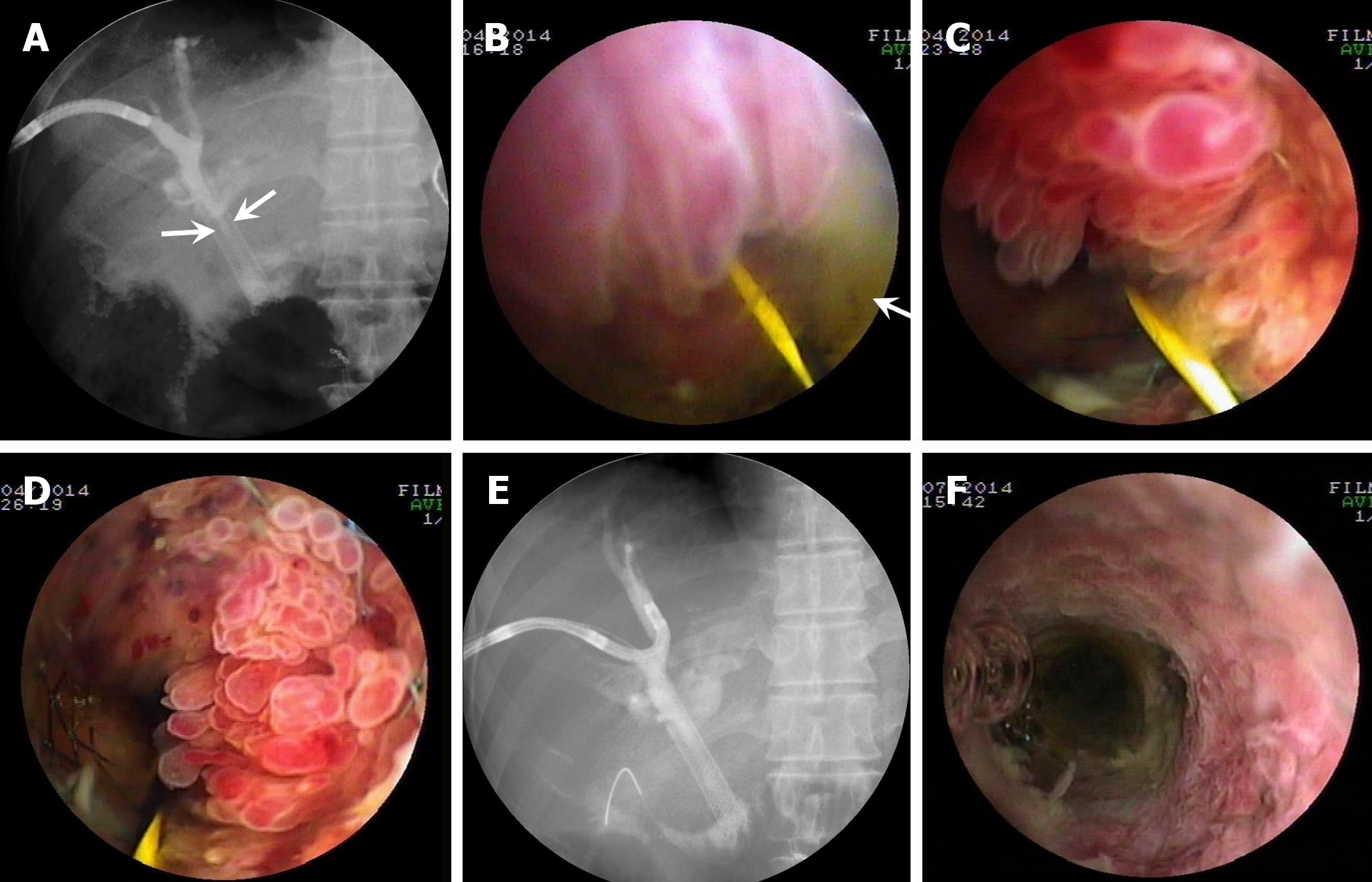
Figure 4 Percutaneous transhepatic cholangioscopy-assisted biliary polypectomy for postoperative recurrent mucin-hypersecreting cast-like type intraductal papillary neoplasm of the bile duct, with therapeutic success in patient four who received transanastomotic metal stenting for intraductal papillary neoplasm of the bile duct one month ago.
A: Cholangiography showing stenosis of the metal stent lumen (arrows); B-D: Percutaneous transhepatic cholangioscopy (PTCS) showed thick mucus (arrow) and multiple villous, frond-like, and fish-egg like protrusions in the stent lumen, respectively; E: Cholangiography showing the improvement of the stricture with a patent metal stent in the lumen; F: Repeated PTCS showing no obvious elevated tumor regrowth in the metal stent 3 months after PTCS-assisted biliary polypectomy.
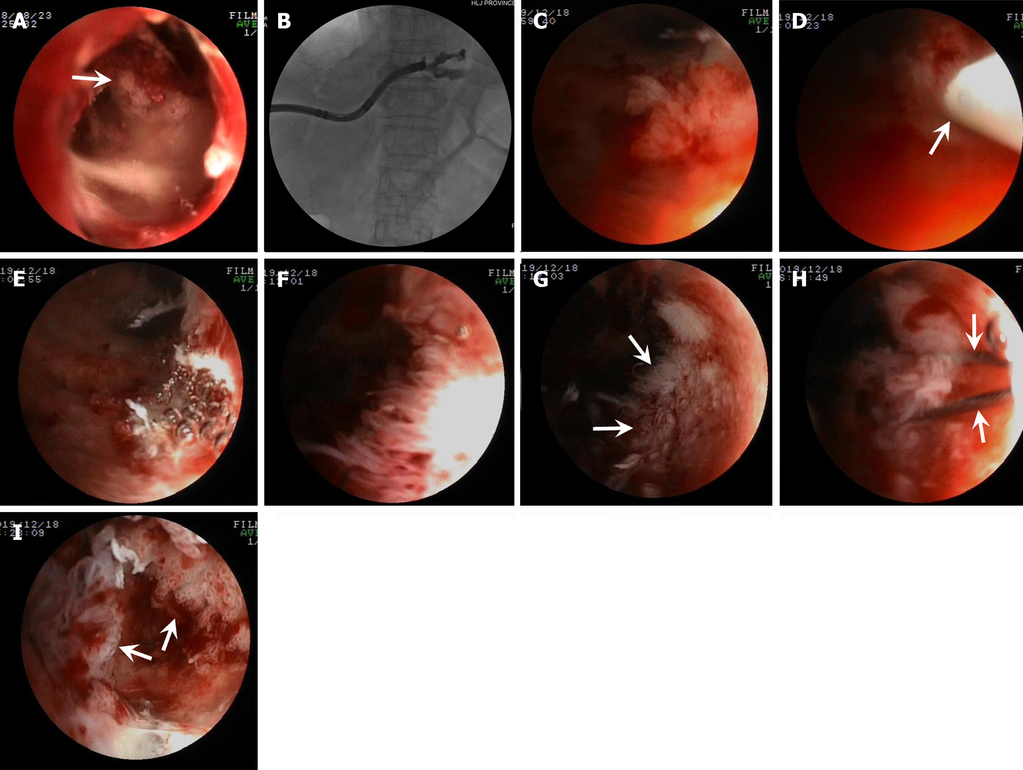
Figure 5 Percutaneous transhepatic cholangioscopy-assisted biliary polypectomy for mucin-hypersecreting cast-like type intraductal papillary neoplasm in the intrahepatic bile duct, with technical success in patient five.
A: Percutaneous transhepatic cholangioscopy (PTCS) showing the biliary tumors at the proximal end of the stricture; B: Cholangiography showing the location of the stricture in the left intrahepatic bile duct; C: PTCS showed a frond-like tumor; D: The snared tumor (the arrow points to the snare sheath); E: The successfully resected tumor with PTCS-assisted biliary polypectomy (PTCS-BP); F: PTCS showing multiple villous growing tumors; G: PTCS showing some residual tumors (arrows) after PTCS-BP; H: Subsequent PTCS-BP performed for the residual villous growing tumors (arrows point to the snare steel wire); I: PTCS showing unresected residual tumors (arrows) in the small ductal lumen.
- Citation: Ren X, Qu YP, Zhu CL, Xu XH, Jiang H, Lu YX, Xue HP. Percutaneous transhepatic cholangioscopy-assisted biliary polypectomy for local palliative treatment of intraductal papillary neoplasm of the bile duct. World J Gastrointest Oncol 2024; 16(5): 1821-1832
- URL: https://www.wjgnet.com/1948-5204/full/v16/i5/1821.htm
- DOI: https://dx.doi.org/10.4251/wjgo.v16.i5.1821


