Copyright
©The Author(s) 2024.
World J Gastrointest Oncol. Mar 15, 2024; 16(3): 933-944
Published online Mar 15, 2024. doi: 10.4251/wjgo.v16.i3.933
Published online Mar 15, 2024. doi: 10.4251/wjgo.v16.i3.933
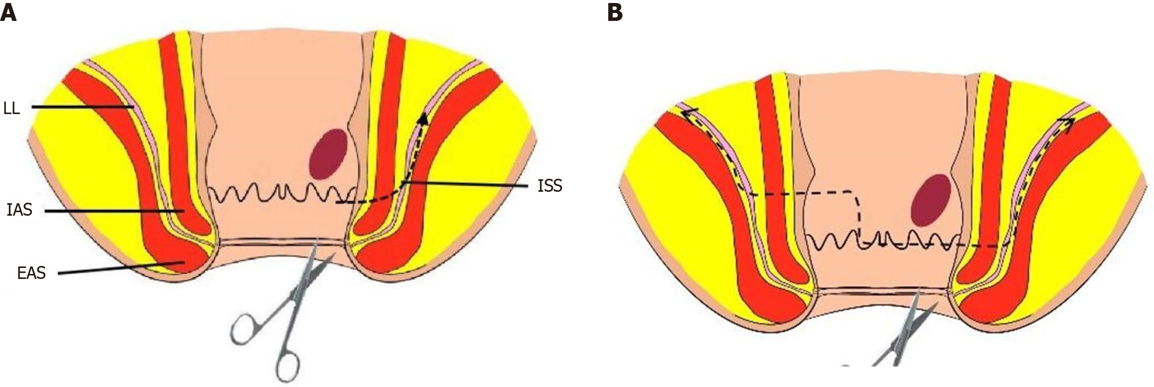
Figure 1 Diagram illustrating the excision range.
A: Surgical resection range of intersphincteric resection (ISR); B: Surgical resection range of modified ISR. EAS: External anal sphincter; IAS: Internal anal sphincter; ISS: Intersphincteric space.
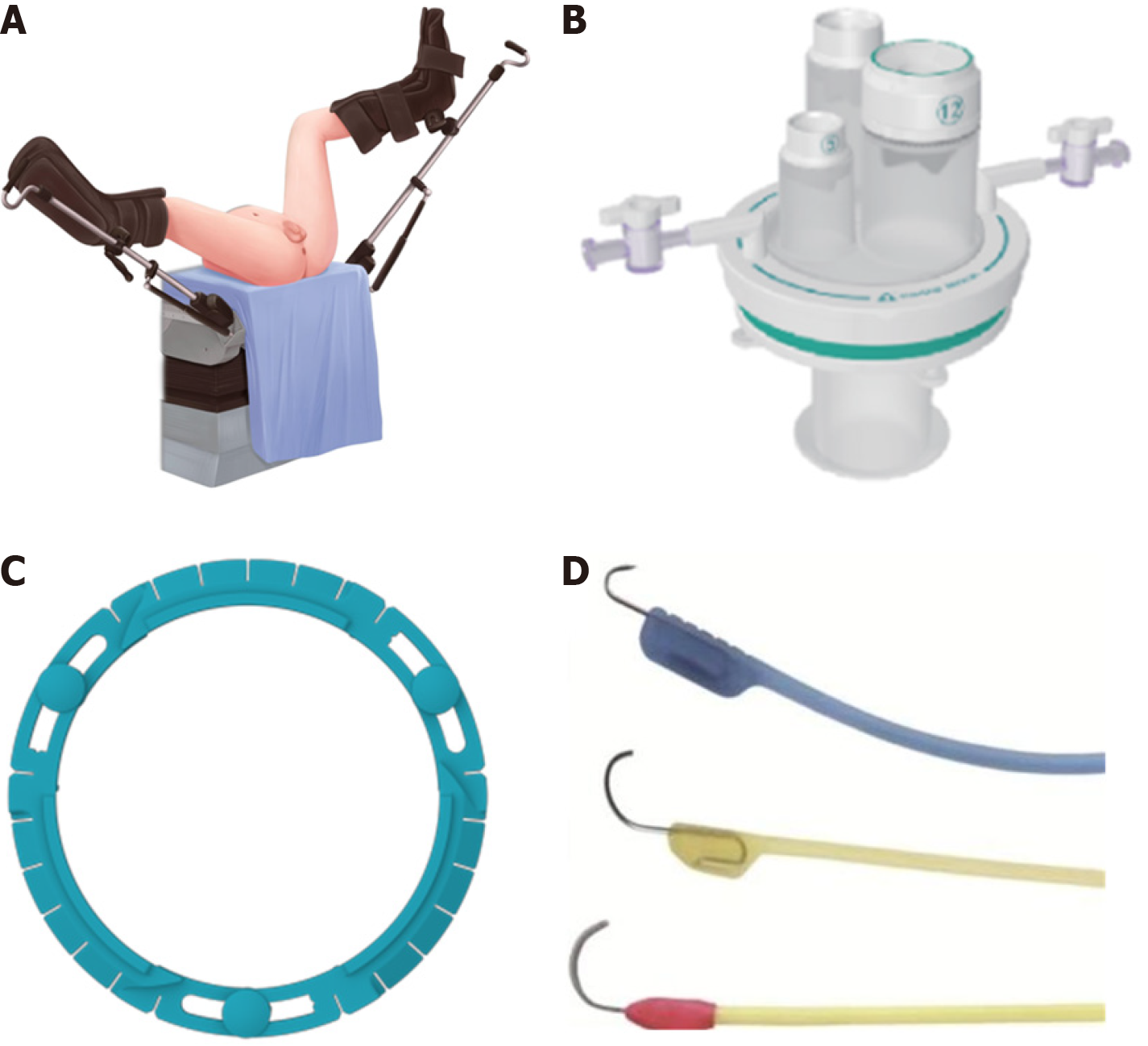
Figure 2 Illustration of patient positioning and equipment.
A: Diagram of the patient's positioning during surgery; B: STAR-PORT soft single-port laparoscopic platform; C and D: Lone star disposable sterile retractor and retraction hooks.
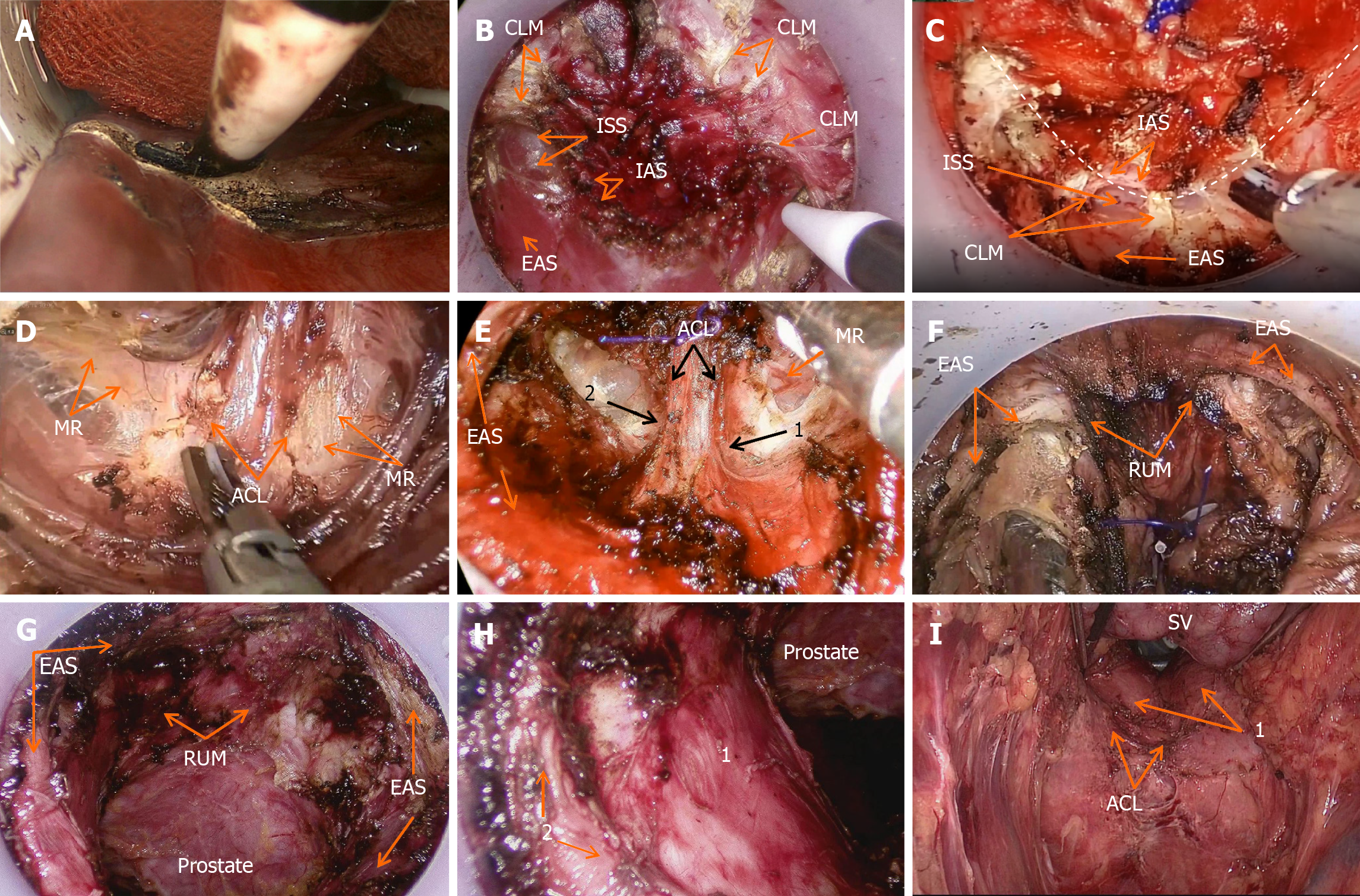
Figure 3 Intraoperative diagrams.
A: Initial exposure of the intersphincteric space; B: Incision to open the radial fibers of the conjoint longitudinal muscle (CLM) and the circular fibers of the internal anal sphincter (IAS); C: "U" shaped dissection of the posterior intersphincteric space (ISS) and the radial fibers of the CLM within it; D: Exposure of the ventral layer of the anococcygeal ligament (ACL) and the distal mesorectum (MR); E: Division of the ventral layer of the ACL (1: Left Hiatal ligament remnant. 2: Right Hiatal ligament remnant); F: External anal sphincter (EAS) and the longitudinal radial fibers of the rectourethralis muscle (RUM); G: Residual end of the RUM and the prostate after transanal endoscopic intersphincteric resection; H: Postoperative view from the anal perspective (1: Complex of the levator ani muscle (LAM) and ESA; 2: Intermediate loop of EAS); I: Postoperative view from the abdominal perspective (1: Complex of the LAM and ESA). SV: Seminal vesicle.
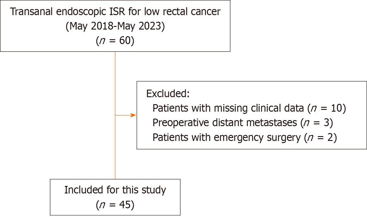
Figure 4 Flowchart of patients included in this study.
ISR: Intersphincteric resection.
- Citation: Xu ZW, Zhu JT, Bai HY, Yu XJ, Hong QQ, You J. Clinical efficacy and pathological outcomes of transanal endoscopic intersphincteric resection for low rectal cancer. World J Gastrointest Oncol 2024; 16(3): 933-944
- URL: https://www.wjgnet.com/1948-5204/full/v16/i3/933.htm
- DOI: https://dx.doi.org/10.4251/wjgo.v16.i3.933


