Copyright
©The Author(s) 2024.
World J Gastrointest Oncol. Mar 15, 2024; 16(3): 857-874
Published online Mar 15, 2024. doi: 10.4251/wjgo.v16.i3.857
Published online Mar 15, 2024. doi: 10.4251/wjgo.v16.i3.857
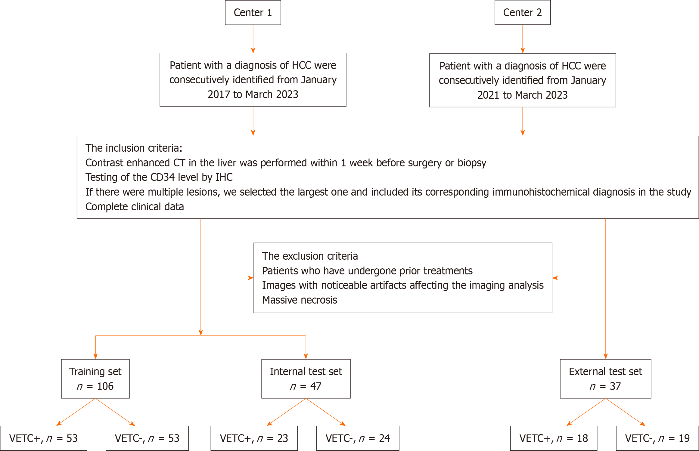
Figure 1 Flow chart of patient recruitment pathway.
HCC: Hepatocellular carcinoma; CT: Computed tomography; IHC: Immunohistochemistry; VETC: Vessels encapsulating tumor clusters; CD34: Cluster of differentiation 34.
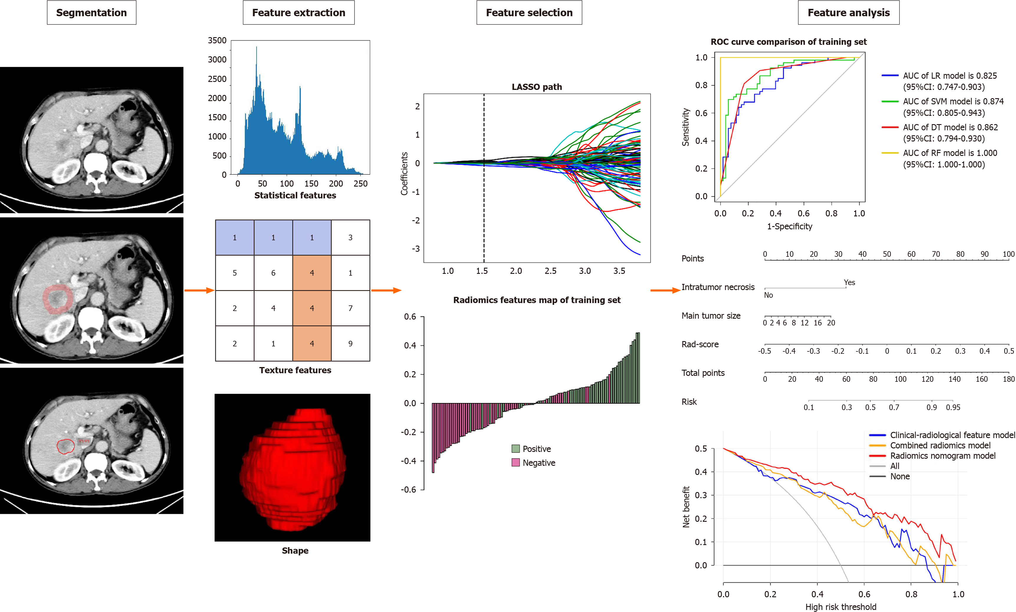
Figure 2 Flowchart of radiomics.
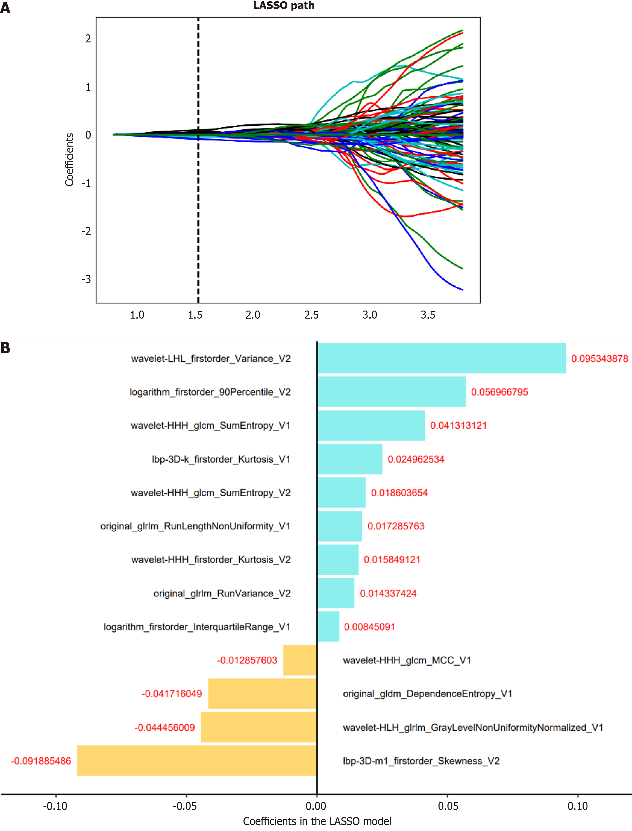
Figure 3 Radiomics feature selection.
A: The least absolute shrinkage and selection operator of the parameterized method was used to select the image omics features by logistic regression; select the optimal alpha of 0.0297 with log(alpha) of -1.527; B: The coefficients of the radiomics features were used for weighting. LASSO: Least absolute shrinkage and selection operator.
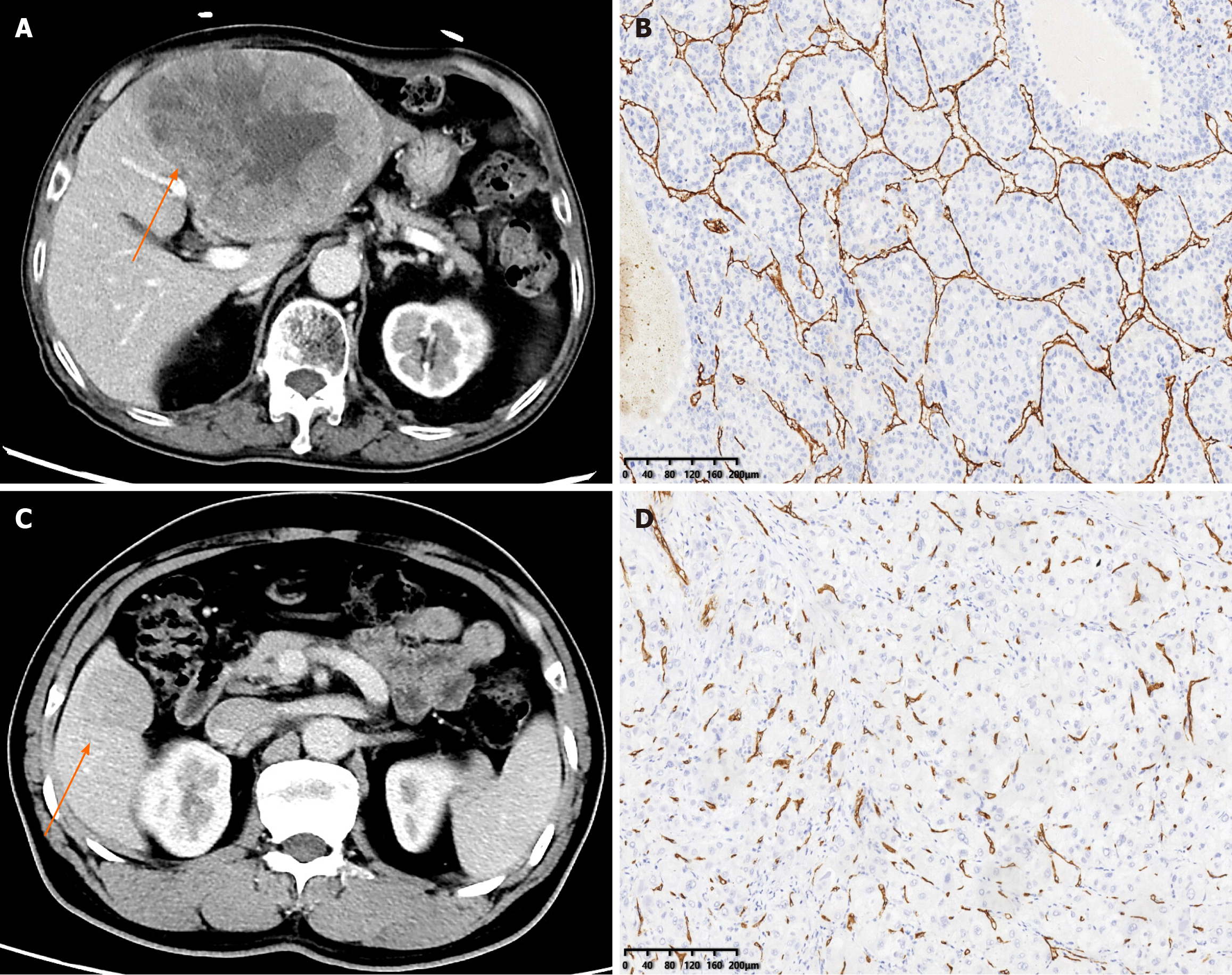
Figure 4 Contrast enhanced computed tomography and immunohistochemical staining for cluster of differentiation 34.
A: Vessels encapsulating tumor cluster (VETC) + hepatocellular carcinoma (HCC) in a 71-year-old man, a mass (the arrow) can be seen in the lateral left lobe of liver; B: Immunohistochemical image for cluster of differentiation 34 (CD34) presented vessels that encapsulated tumor clusters and formed cobweb-like networks (original magnification, × 100); C: VETC-HCC in a 52-year-old man, a mass (the arrow) can be seen in the anterior right lobe of the liver; D: Immunohistochemical image for CD34 presented vessels with discrete lumens (original magnification, × 100).
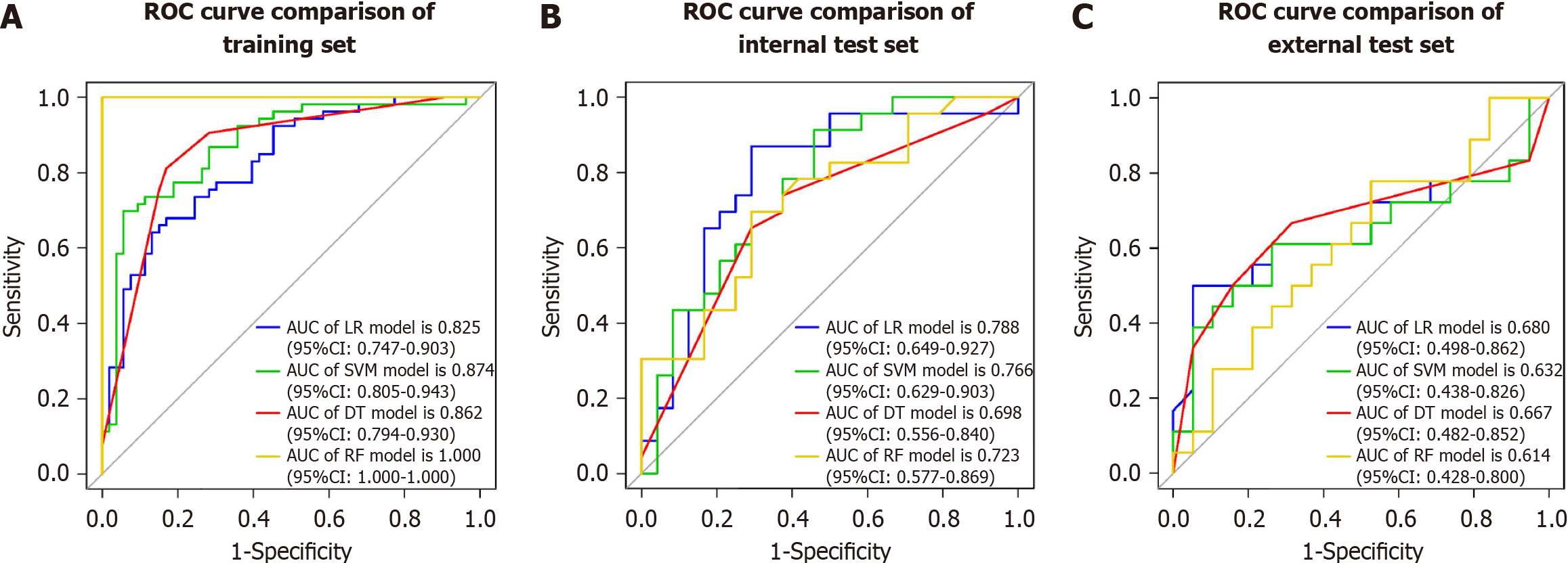
Figure 5 Receiver operating characteristic of the combined radiomics models.
A: Receiver operating characteristic (ROC) of combined radiomics model using four machine learning algorithms on the training set; B: ROC of combined radiomics model using four machine learning algorithms on the internal test set; C: ROC of combined radiomics model using four machine learning algorithms on the external test set. ROC: Receiver operating characteristic; LR: Logistic regression; SVM: Support vector machine; DT: Decision tree; RF: Random forest; CI: Confidence interval; AUC: Area under the curve.
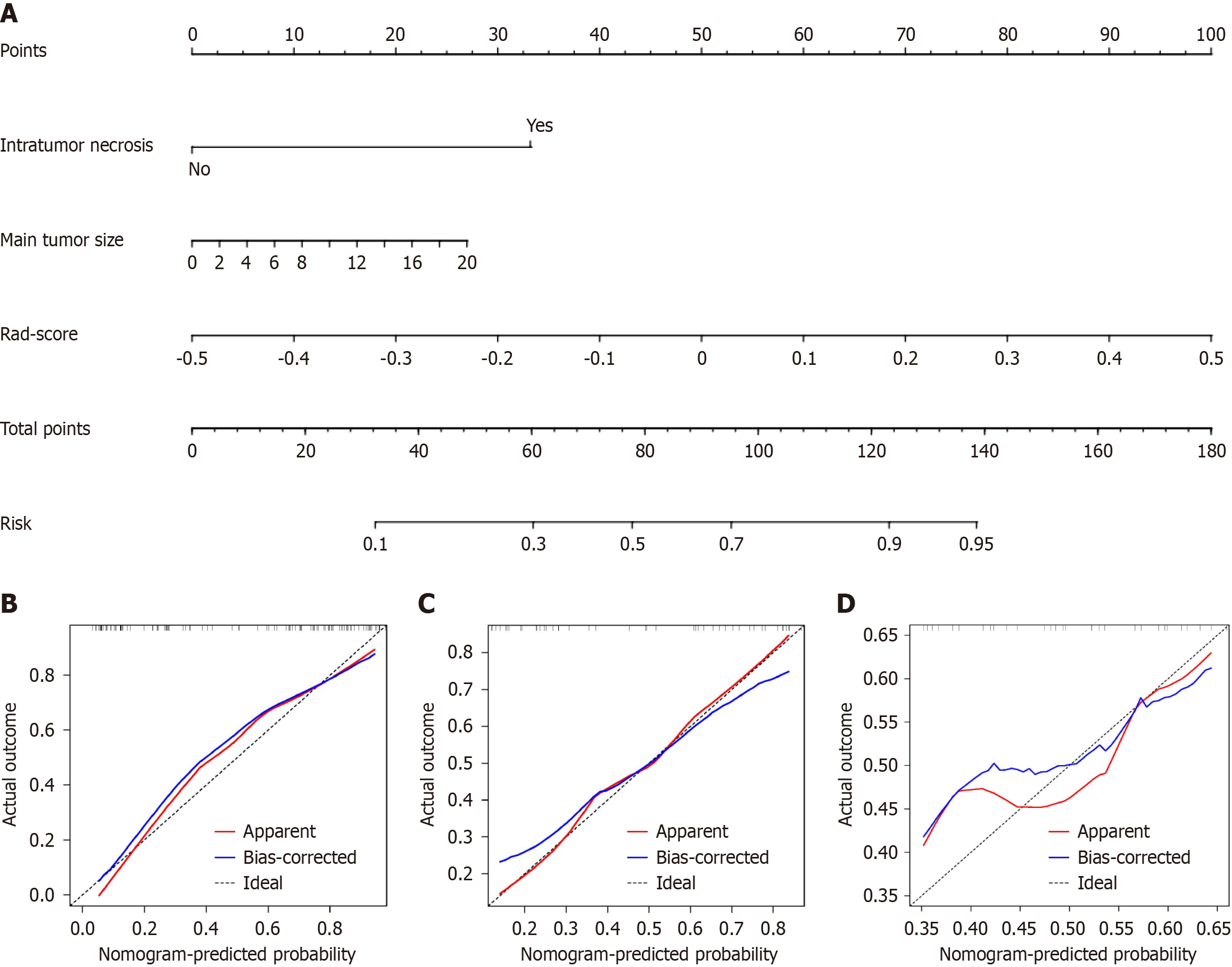
Figure 6 The radiomics nomogram and calibration curves for the radiomics nomogram.
A: The radiomics nomogram combining intratumor necrosis, main tumor size, and radiomics score, was developed on the training set; B: Calibration curves for the radiomics nomogram on the training set; C: Calibration curves for the radiomics nomogram on the internal test set; D: Calibration curves for the radiomics nomogram on the external test set.
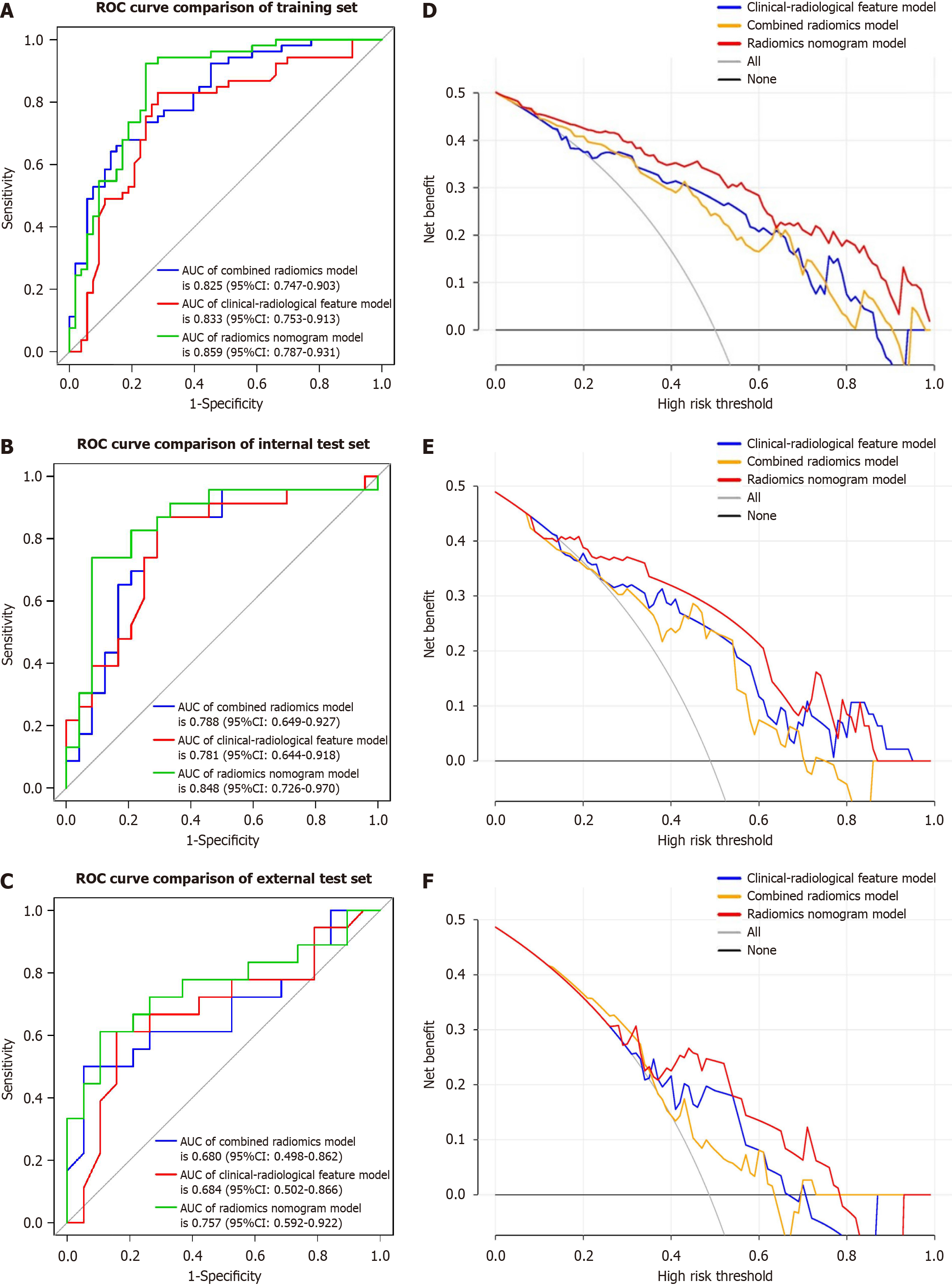
Figure 7 Receiver operating characteristic and decision curve analysis for three models.
A: Receiver operating characteristic (ROC) of clinical-radiological features on the training set; B: ROC of clinical-radiological features on the internal test set; C: ROC of clinical-radiological features on the external test set; D: Decision curve analysis on the training set; E: Decision curve analysis on the internal test set; F: Decision curve anal. ROC: Receiver operating characteristic; CI: Confidence interval; AUC: Area under the curve.
- Citation: Zhang C, Zhong H, Zhao F, Ma ZY, Dai ZJ, Pang GD. Preoperatively predicting vessels encapsulating tumor clusters in hepatocellular carcinoma: Machine learning model based on contrast-enhanced computed tomography. World J Gastrointest Oncol 2024; 16(3): 857-874
- URL: https://www.wjgnet.com/1948-5204/full/v16/i3/857.htm
- DOI: https://dx.doi.org/10.4251/wjgo.v16.i3.857


