Copyright
©The Author(s) 2024.
World J Gastrointest Oncol. Nov 15, 2024; 16(11): 4506-4513
Published online Nov 15, 2024. doi: 10.4251/wjgo.v16.i11.4506
Published online Nov 15, 2024. doi: 10.4251/wjgo.v16.i11.4506
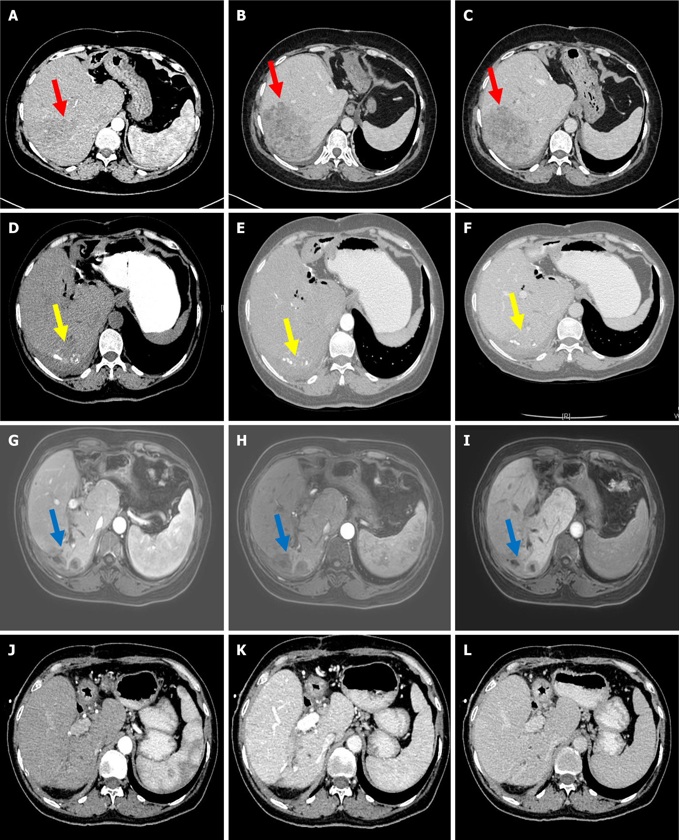
Figure 1 Radiological images of the patient before and after conversion therapy.
A-C: computed tomography (CT) examination shows that the tumor is in the right lobe of the liver before conversion therapy; D-F: CT and magnetic resonance imaging; G-I: Examinations both indicate a decreased tumor in the right lobe of the liver before surgery; J-L: Seven months after surgery, no recurrence or metastasis was observed under the CT. The red, yellow, and blue arrows all head towards the tumor.
Figure 2 Images of HE and immunohistochemical staining of the biopsy specimen (× 200).
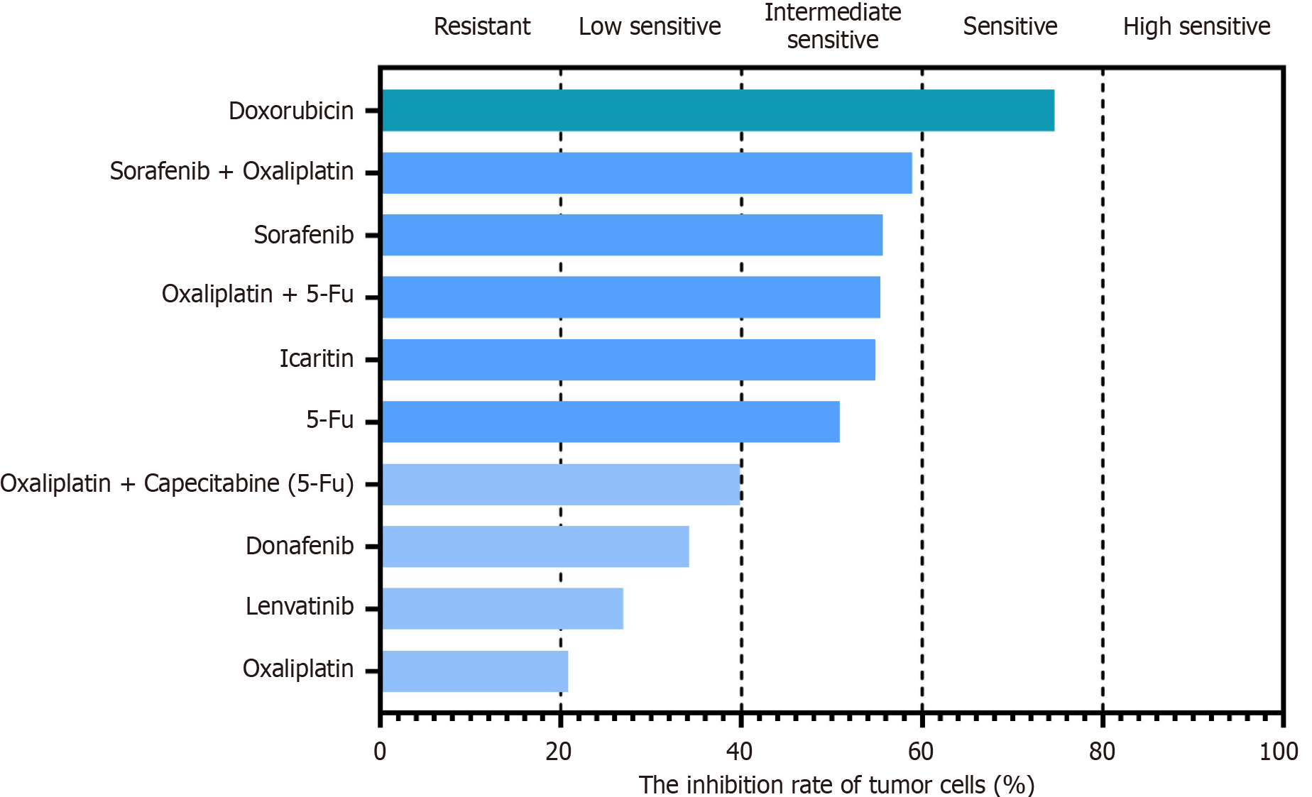
Figure 3 The drug sensitivity results of the patient-derived organoid from hepatocellular carcinoma.
- Citation: He YG, Wang Z, Li J, Xi W, Zhao CY, Huang XB, Zheng L. Pathologic complete response to conversion therapy in hepatocellular carcinoma using patient-derived organoids: A case report. World J Gastrointest Oncol 2024; 16(11): 4506-4513
- URL: https://www.wjgnet.com/1948-5204/full/v16/i11/4506.htm
- DOI: https://dx.doi.org/10.4251/wjgo.v16.i11.4506


