Copyright
©The Author(s) 2024.
World J Gastrointest Oncol. Nov 15, 2024; 16(11): 4436-4455
Published online Nov 15, 2024. doi: 10.4251/wjgo.v16.i11.4436
Published online Nov 15, 2024. doi: 10.4251/wjgo.v16.i11.4436
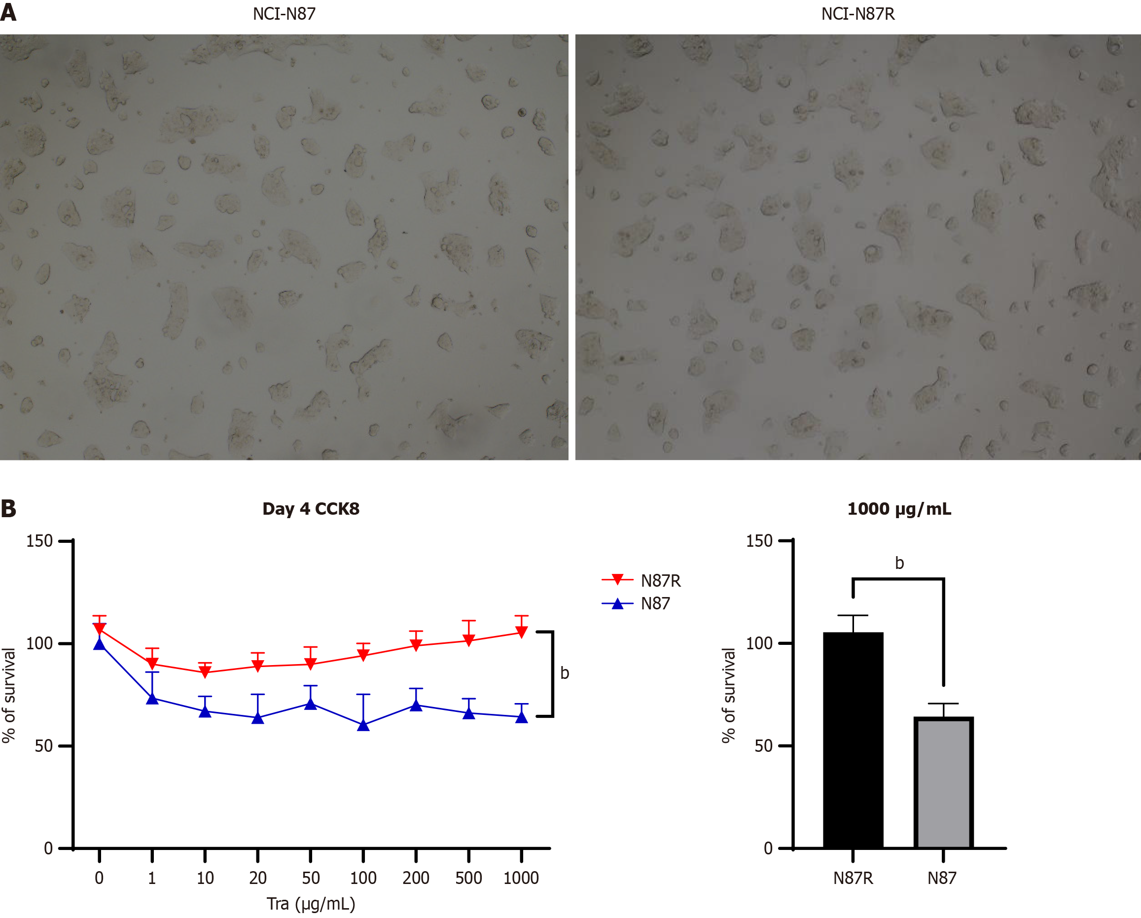
Figure 1 Construction of trastuzul tumor resistance model.
A: The morphology of the resistant strain NCI-N87R is similar to that of the parent cell line NCI-N87; B: Cells were treated with 1000 μg/mL trastuzumab for 4 days, and cell viabilities were detected by cell counting kit-8 assay. bP < 0.0001. CCK8: Cell counting kit-8; Tra: Trastuzumab.
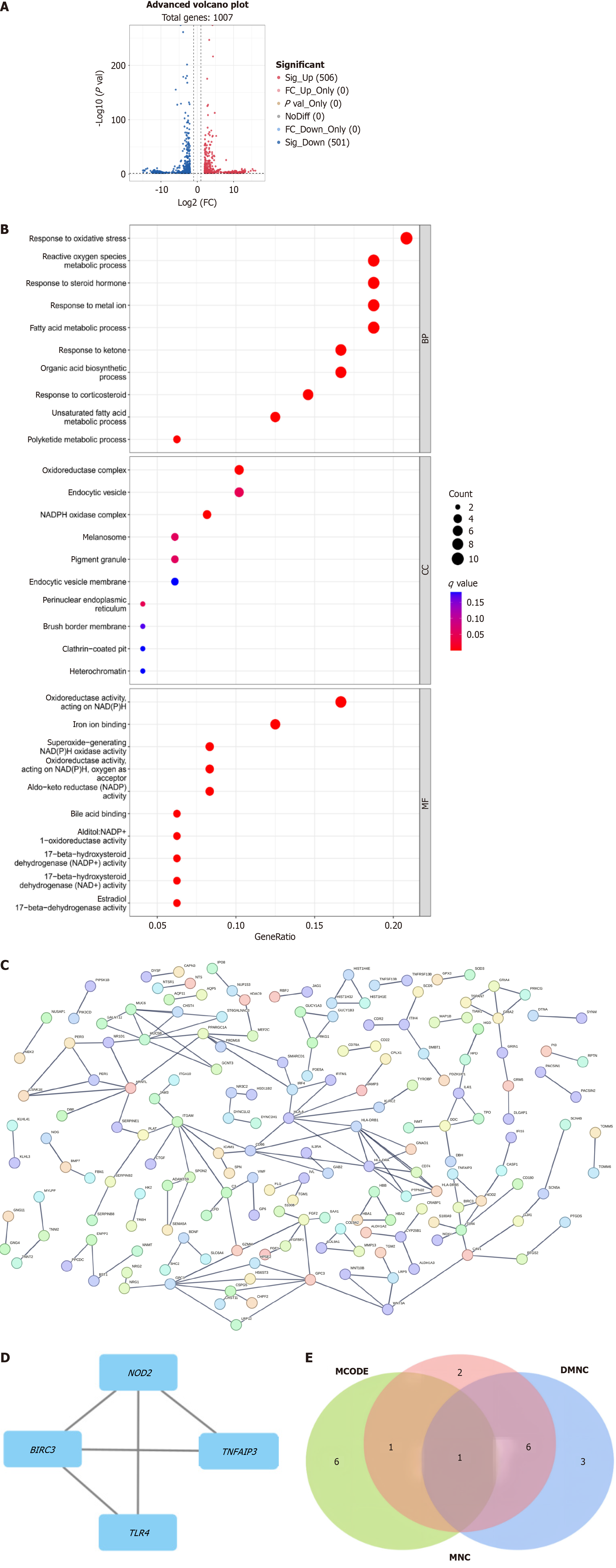
Figure 2 High throughput sequencing and bioinformatics analysis screening Hub genes.
A: The volcano plot was generated from mRNA sequencing analysis of NCI-N87 and NCI-N87R; B: Gene pathway analysis was performed; C: Protein interaction network analysis for BIRC3 was performed using the STRING online tools; D: the top differentially expressed genes were identified by the maximum neighborhood component centrality and neighborhood component centrality algorithms; E: Venn diagram was presented to show the top 10 genes selected using different algorithms, based on the CytoHubba online tools. FC: Fold change; DMNC: Density of maximum neighborhood component; MNC: Maximum neighborhood component.
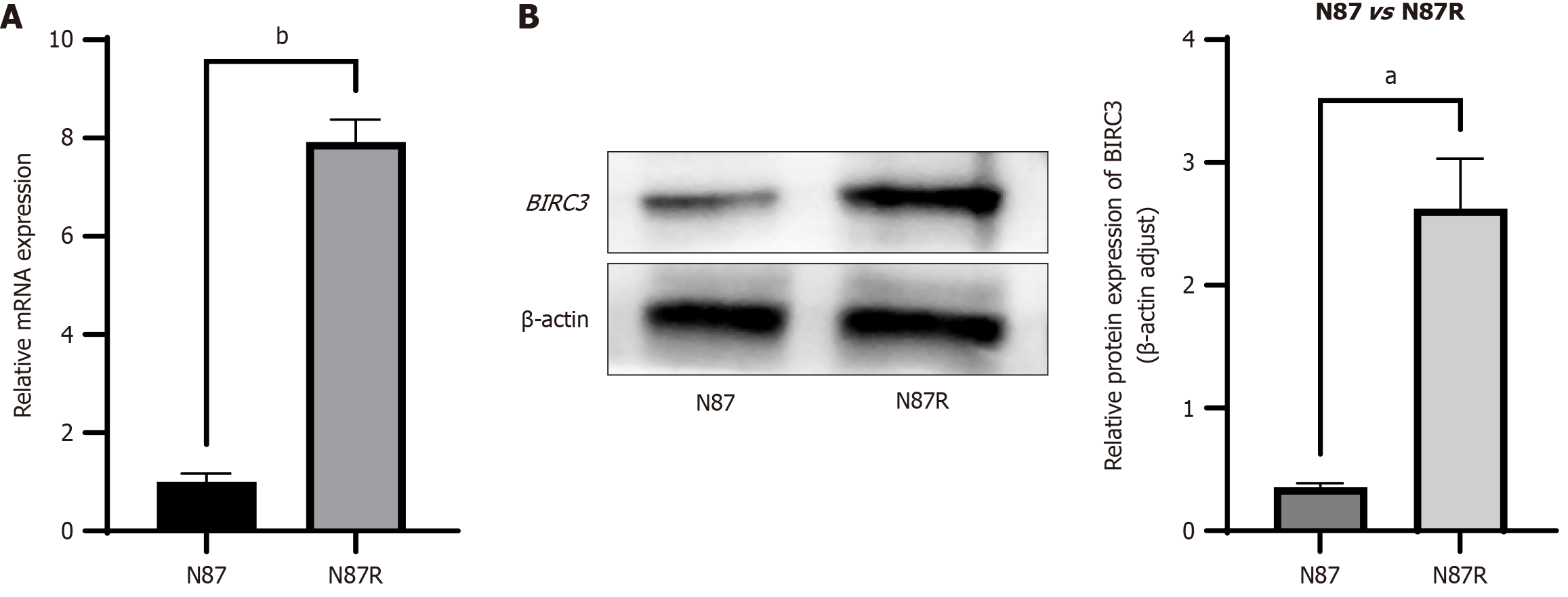
Figure 3 Expression of BIRC3 in human gastric cancer cell lines and trastuzul resistant cell lines.
A: mRNA levels of BIRC3 in NCI-N87, and NCI-N87R cells were examined by reverse transcription polymerase chain reaction; GAPDH was used as a control; B: Protein levels of BIRC3 in NCI-N87, and NCI-N87R cells were examined by western blotting, β-actin was a negative control. aP < 0.001; bP < 0.0001.
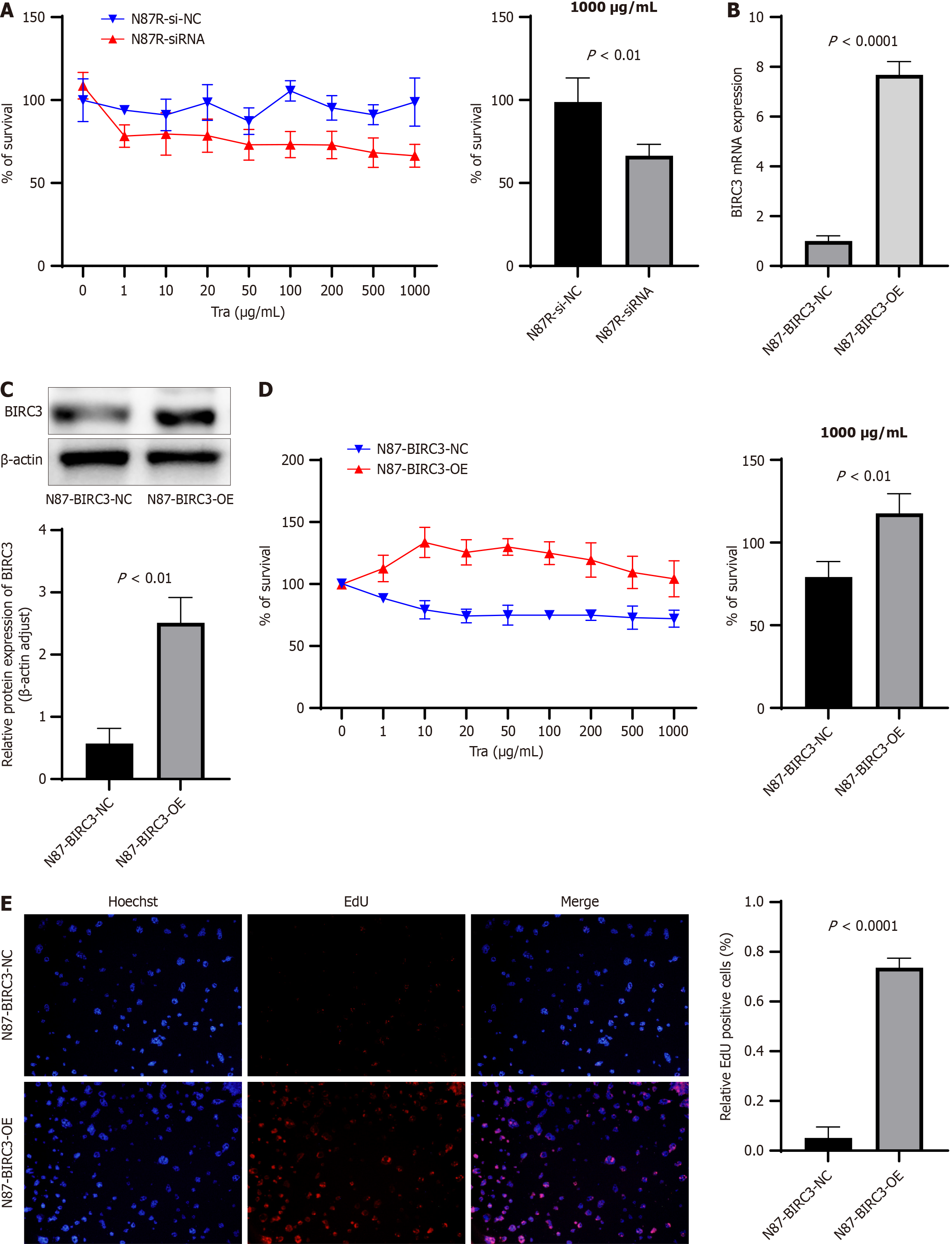
Figure 4 BIRC3 promotes the resistance of human epidermal growth factor receptor 2-position gastric cancer to trastuzumab by promote the proliferation of tumor cell.
A: BIRC3 was depleted using BIRC3-siRNA in NCI-N87R cells. Cell viabilities of N87R-siRNA cells and N87R-siNC 1000 μg/mL trastuzumab were examined by cell counting kit-8 assay; B: mRNA levels of BIRC3 in N87 cells after transfection of BIRC3 overexpressing plasmids or control empty plasmids (designated as N87-BIRC3-OE and N87-BIRC3-NC) were examined by reverse transcription polymerase chain reaction; C: Protein levels of BIRC3 in N87-BIRC3-OE and N87-BIRC3-NC were examined by western blotting. β-actin was a negative control; D: Cell viabilities of N87-BIRC3-OE and N87-BIRC3-NC cells 1000 μg/mL trastuzumab were examined by cell counting kit-8 assay; E: Cell viabilities of N87-BIRC3-OE and N87-BIRC3-NC cells 1000 μg/mL trastuzumab were examined by 5-ethynyl-2’-deoxyuridine assay. Tra: Trastuzumab; EdU: 5-ethynyl-2’-deoxyuridine.
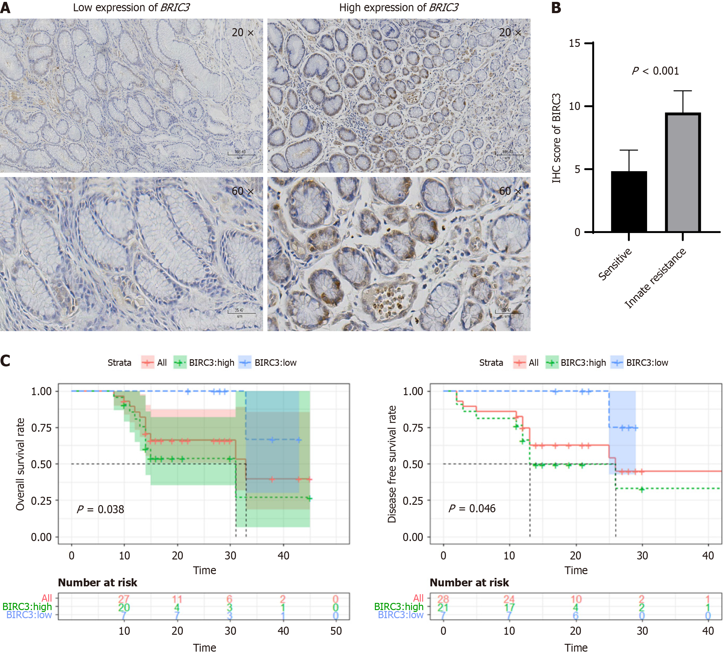
Figure 5 BIRC3 is correlated with trastuzamab resistance and poor prognosis and the in human epidermal growth factor receptor 2-positive gastric cancer patients.
A: Immunohistochemistry (IHC) staining for BIRC3 in gastric cancer (GC) tissues were shown; B: 28 human epidermal growth factor receptor 2 (HER2)-positive patients were stratified into negative and positive groups according to IHC staining for BIRC3; C: The IHC results demonstrated significantly higher expression levels of BIRC3 in the trastuzumab resistant group compared to the sensitive group. Kaplan-Meier analysis of overall survival in HER2-positive GC patients was performed based on BIRC3 levels. IHC: Immunohistochemistry.
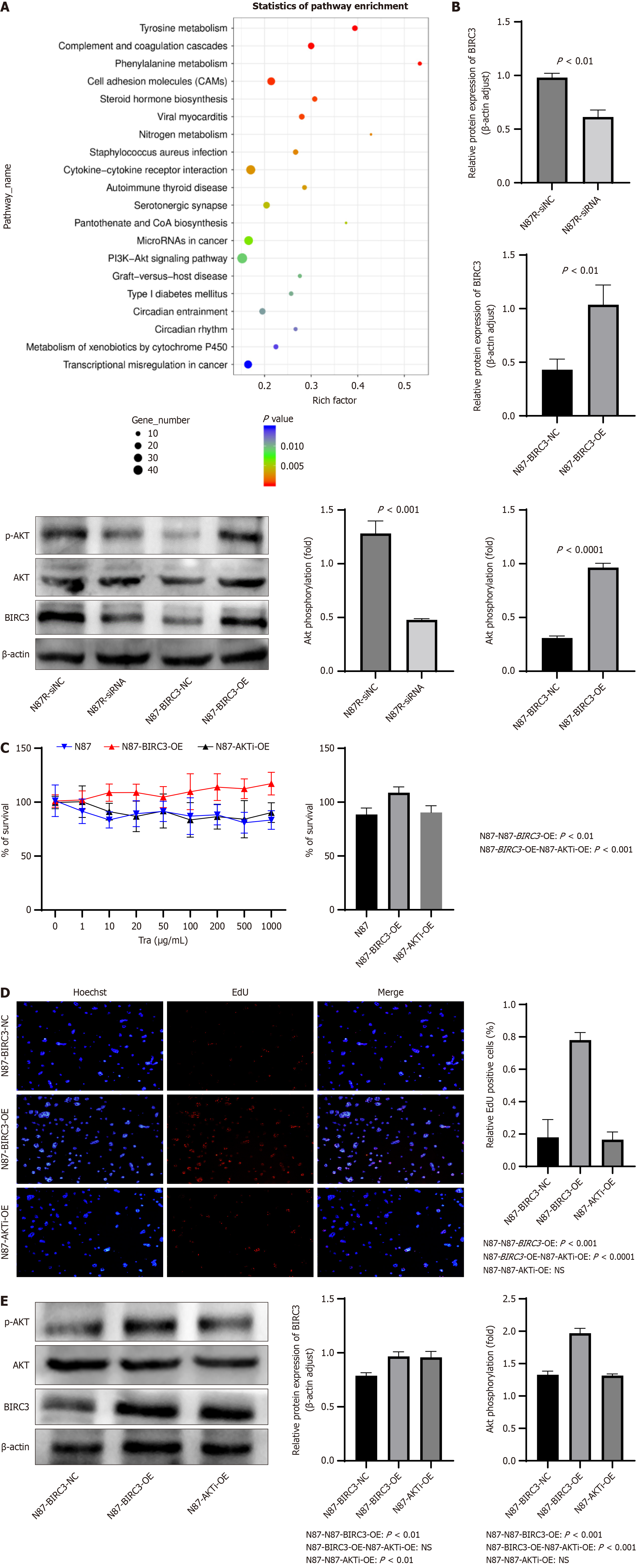
Figure 6 Tumor cell-derived BIRC3 enhancing the proliferation of human epidermal growth factor receptor 2-positive gastric cancer cell treated with trastuzumab activating AKT pathway.
BIRC3 siRNA or siRNA-NC was transfected into NCI-N87R cells (designated as N87R-siRNA and N87R-siNC). NCI-N87 cells were pretreated with an Akt inhibitor (MK-2206) and transfected with the overexpression plasmid pLVX-IRES-puro-BIRC3 (designated as N87-AKTi-OE). A: Revealed that the differential genes were mainly enriched in pathways by the Kyoto encyclopedia of genes and genomes enrichment analysis; B: Protein levels of BIRC3 and AKT, p-AKT were examined by western blotting. β-actin was a negative control; C: Cell viabilities of N87, N87-BIRC3-OE and N87-BIRC3-NC cells 1000μg/mL trastuzumab were examined by cell counting kit-8 assay; D: Cell viabilities of N87, N87-BIRC3-OE and N87-BIRC3-NC cells 1000μg/mL trastuzumab were examined by 5-ethynyl-2’-deoxyuridine assay; E: Protein levels of BIRC3 and AKT, p-AKT in N87,N87-BIRC3-OE, and N87-AKTi-OE cells were examined by western blotting. β-actin was a negative control. Tra: Trastuzumab; EdU: 5-ethynyl-2’-deoxyuridine; NS: No significance.
Figure 7 Tumor cell-derived BIRC3 inhibits apoptosis by activating AKT pathway, lead to drug resistance to trastuzumab.
A: Experimental in Balb/c nude mice. Tumor volume images, tumor weight and tumor growth curves of each group were shown. The different treatment groups included: (1) The stable control NCI-N87 cells (N87 group); (2) The stable BIRC3 overexpressing NCI-N87 cells with the trastuzumab treatment (N87-LV-BIRC3 group); and (3) The NCI-N87 transfected with the overexpressed virus LV-GFP-puro-BIRC3 and treated with AKT inhibitor (MK-2206) (N87-LV-BIRC3-AKTi group); B: (1) The NCI-N87 was developed into a trastuzumab-resistant cell line(N87R group); (2) The group inoculated with NCI-N87R cells transfected with the knockout virus LV-GFP-puro-BIRC3-si (N87-LV-BIRC3 group); C and D: Immunohistochemistry staining for BIRC3, pAKT and caspase 3 was presented, and the immunohistochemistry scores for BIRC3, pAKT and caspase 3 were statistically analyzed. IHC: Immunohistochemistry; HE: Hematoxylin-eosin; NS: No significance.
- Citation: Li SL, Wang PY, Jia YP, Zhang ZX, He HY, Chen PY, Liu X, Liu B, Lu L, Fu WH. BIRC3 induces the phosphoinositide 3-kinase-Akt pathway activation to promote trastuzumab resistance in human epidermal growth factor receptor 2-positive gastric cancer. World J Gastrointest Oncol 2024; 16(11): 4436-4455
- URL: https://www.wjgnet.com/1948-5204/full/v16/i11/4436.htm
- DOI: https://dx.doi.org/10.4251/wjgo.v16.i11.4436


