Copyright
©The Author(s) 2024.
World J Gastrointest Oncol. Oct 15, 2024; 16(10): 4281-4288
Published online Oct 15, 2024. doi: 10.4251/wjgo.v16.i10.4281
Published online Oct 15, 2024. doi: 10.4251/wjgo.v16.i10.4281
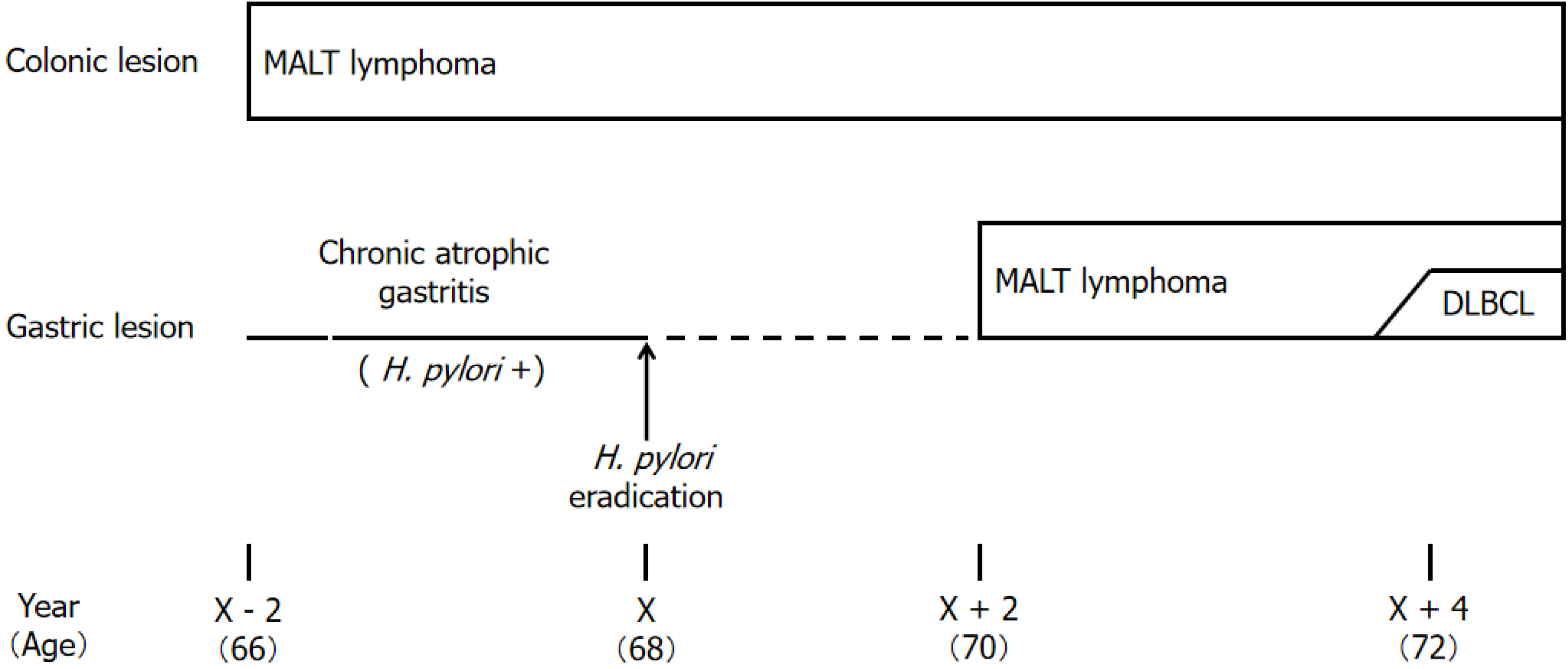
Figure 1 Clinical course chart.
MALT: Mucosa-associated lymphoid tissue; DLBCL: Diffuse large B-cell lymphoma.
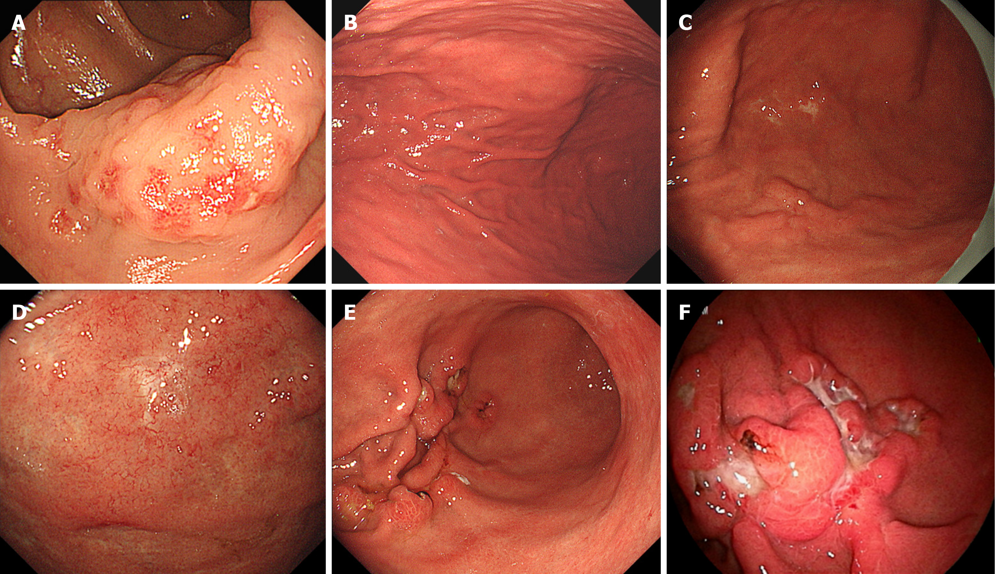
Figure 2 Endoscopic findings.
Colonoscopy image (A) and esophagogastroduodenoscopy image (B-F). A: This image was taken at the time of discovery of colonic mucosa-associated lymphoid tissue (MALT) lymphoma (at the age of 66); B: This image was taken at the time of the discovery of chronic atrophic gastritis associated with Helicobacter pylori (H. pylori) infection (at the age of 68); C: MALT lymphoma was detected in the greater curvature of the body of the stomach 2 years and 9 months after H. pylori eradication (at the age of 70); D: MALT lymphoma was found in the fundus of the stomach 3 years and 3 months after the eradication of H. pylori (at the age of 71); E: Four years and three months after the eradication of H. pylori (at the age of 72), the lymphoma in the gastric body had progressed and transformed into diffuse large B-cell lymphoma; F: At the time of admission to our department, the lymphoma lesions had further progressed.
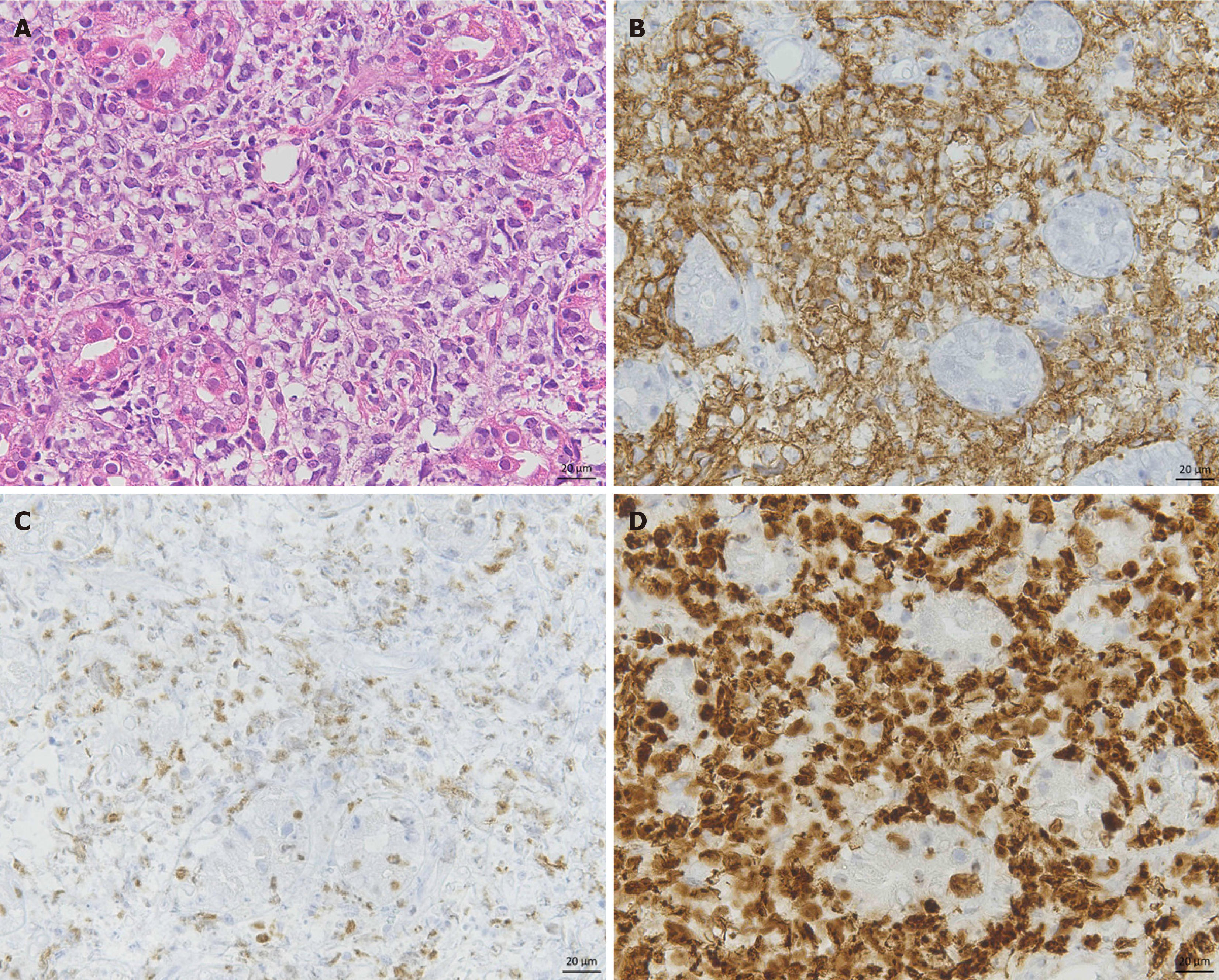
Figure 3 Histopathological findings.
A: Large abnormal lymphoid cells with enlarged nuclei were proliferating into the glandular ducts (Hematoxylin & eosin staining); B and C: These lymphoid tumor cells were positive for CD20 immunostaining (B) and for c-myc immunostaining (C); D: The MIB-1 index was positive in more than 90% of the tumor cells. Pathological diagnosis of diffuse large B-cell lymphoma was made.
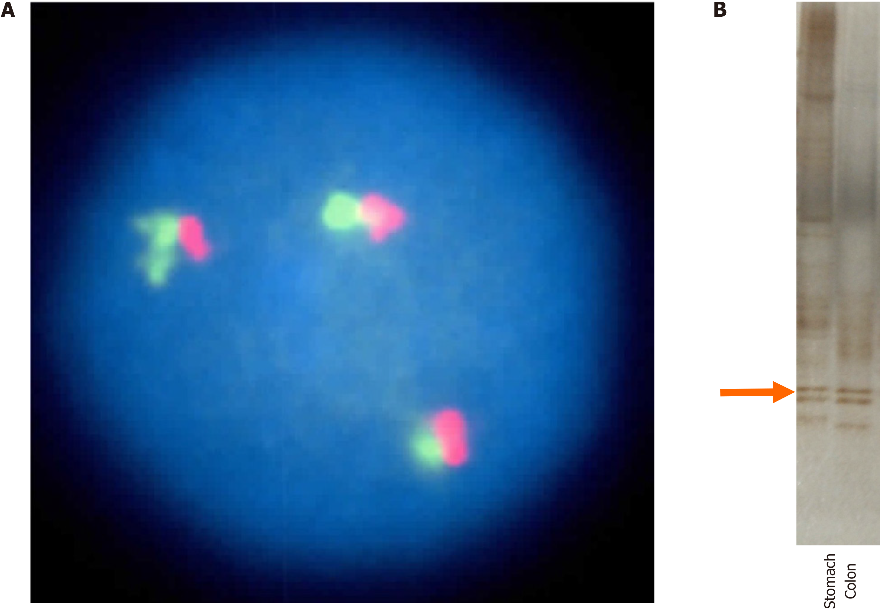
Figure 4 Molecular biological analysis.
A: Three copies of the MALT1 gene were detected in the nuclei of lymphoma cells (Fluorescence in situ hybridization); B: The bands indicating monoclonality (orange arrows) matched, revealing that the colonic and gastric lesions had the same clonality (polymerase chain reaction-based single-strand conformational polymorphism).
- Citation: Saito M, Tanei ZI, Tsuda M, Suzuki T, Yokoyama E, Kanaya M, Izumiyama K, Mori A, Morioka M, Kondo T. Transformed gastric mucosa-associated lymphoid tissue lymphoma originating in the colon and developing metachronously after Helicobacter pylori eradication: A case report. World J Gastrointest Oncol 2024; 16(10): 4281-4288
- URL: https://www.wjgnet.com/1948-5204/full/v16/i10/4281.htm
- DOI: https://dx.doi.org/10.4251/wjgo.v16.i10.4281


