Copyright
©The Author(s) 2024.
World J Gastrointest Oncol. Oct 15, 2024; 16(10): 4232-4243
Published online Oct 15, 2024. doi: 10.4251/wjgo.v16.i10.4232
Published online Oct 15, 2024. doi: 10.4251/wjgo.v16.i10.4232
Figure 1 Microbiomes presented in hepatocellular carcinoma tissue.
Fluorescence in situ hybridization (FISH) was performed on slides of human hepatocellular carcinoma (HCC) and normal liver tissue. A: Fluorescence microscopy was used to analyze FISH signals in HCC; B: FISH signal in normal liver tissue. The image on the left depicts DAPI staining, while the image on the right illustrates EUB338 probe staining.
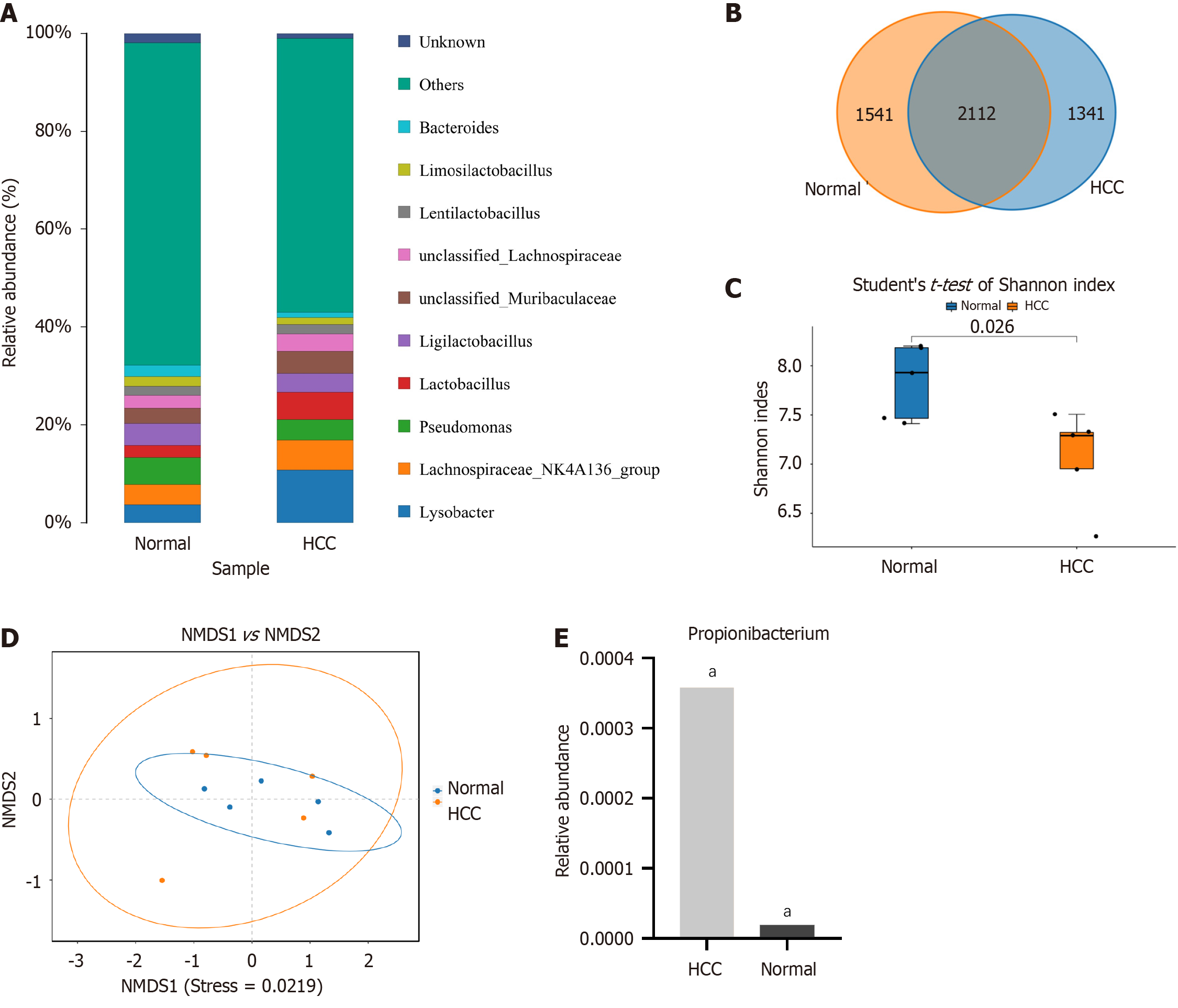
Figure 2 Heterogeneous microbiome in hepatocellular carcinoma and peritumoral tissue.
A: The abundance of microbiomes in hepatocellular carcinoma (HCC) and peritumoral tissues ranked among the top 10 at the generic level; B: Venn diagram of operational classification units composition in HCC and peritumoral tissues; C: Shannon index of response alpha diversity; D: The β diversity exhibited substantial disparities between the HCC and peritumoral groups; E: Relative content of Propionibacterium in HCC and peritumoral tissues. (aP < 0.001). HCC: Hepatocellular carcinoma.
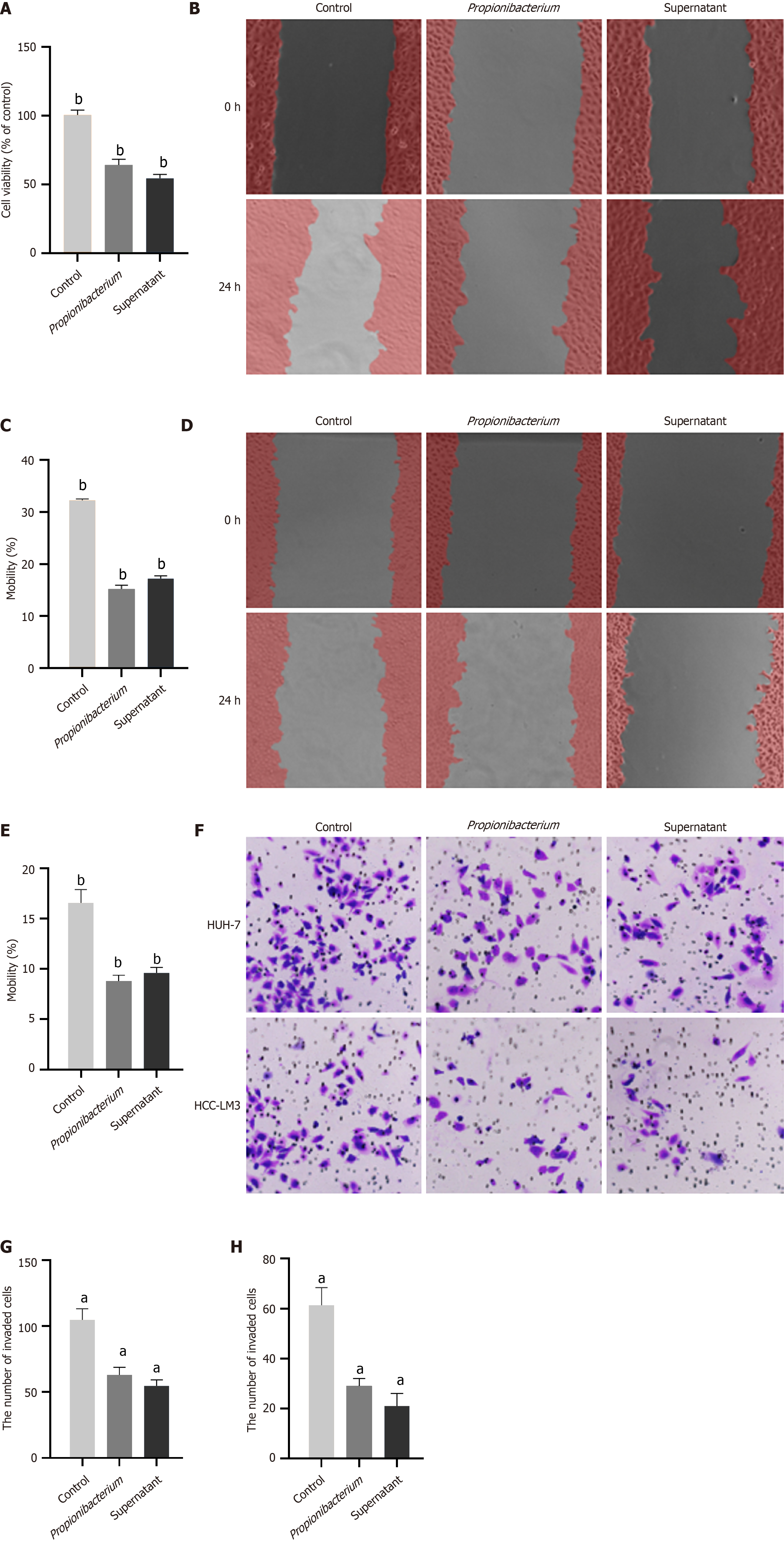
Figure 3 Propionibacterium inhibited the proliferation and migration of hepatocellular carcinoma cells.
A: Cell viability was measured through CCK-8 assay. HUH-7 cells were exposed to Propionibacterium and its supernatant; B: The migration of HUH-7 cells in control, Propionibacterium and supernatant groups was evaluated by wound healing assay; C: Changes in mobility of HUH-7 cells; D: Results of 24 hours wound healing assay on hepatocellular carcinoma (HCC)-LM3 cells; E: Changes in migration rate of HCC-LM3 cells; F: Transwell assay of HUH-7 and HCC-LM3 cells. The migrating cells were observed using a 40 × microscope; G: The quantity of migrated HUH-7 cells; H: The quantity of migrated HCC-LM3 cells. aP < 0.0001, bP < 0.001.
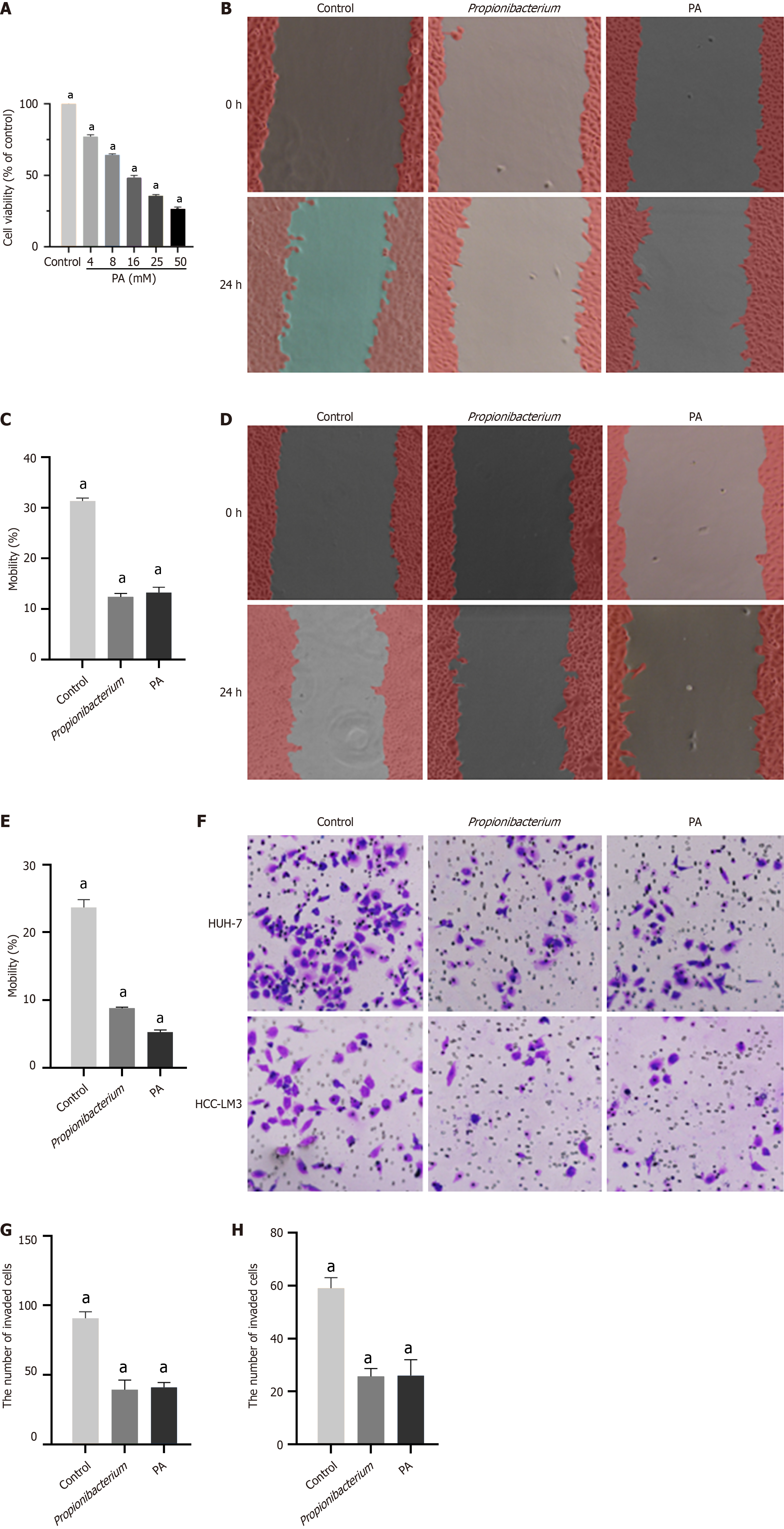
Figure 4 Propionic acid played an inhibitory role as a metabolite of Propionibacterium.
A: Effects of different concentrations of propionic acid (PA) on the cell viability of HUH-7 cells; B: Wound healing assay results of HUH7 cells in control, Propionibacterium and PA group; C: Changes in mobility of HUH-7 cells; D: Results of wound healing assay on hepatocellular carcinoma (HCC)-LM3 cells; E: The mobility of HCC-LM3 cells in Propionibacterium and PA groups decreased significantly; F: Transwell assay results of HUH7 and HCC-LM3 cells; G: The quantity of migrated HUH-7 cells; H: The quantity of migrated HCC-LM3 cells. aP < 0.0001. PA: Propionic acid.
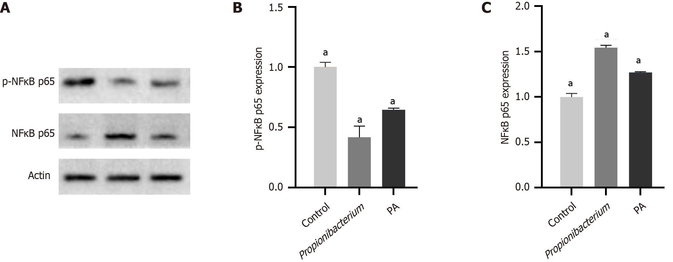
Figure 5 Propionibacterium and propionic acid inhibited the NF-κB pathway.
A: Western blot was performed after coculture of Propionibacterium and propionic acid (PA) with HUH-7 cells; B: Relative expression level of p-NF-κB p65. The expression of p-NF-κB p65 was significantly decreased in Propionibacterium and PA groups; C: Relative expression level of NF-κB p65. aP < 0.0001. PA: Propionic acid.
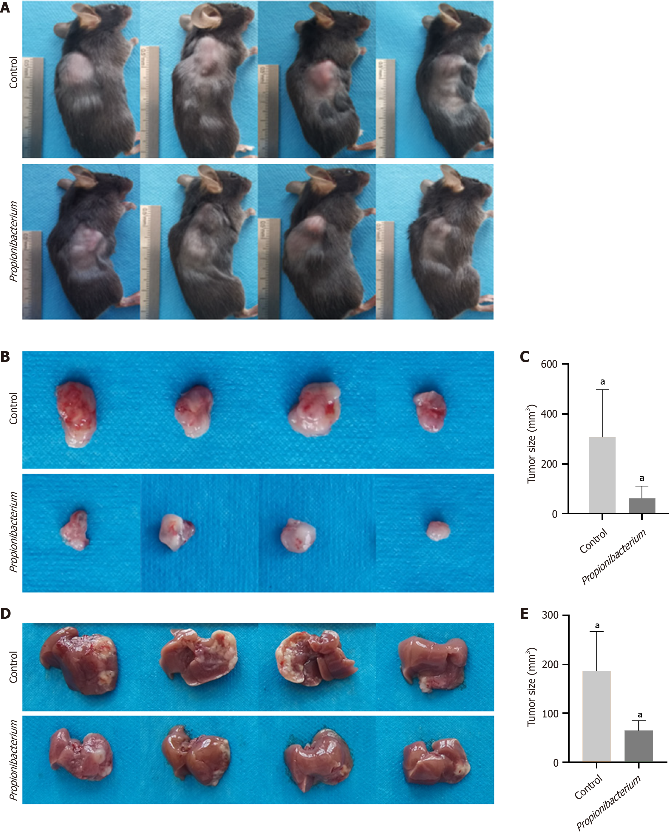
Figure 6 Propionibacterium and propionic acid repressed in vivo hepatocellular carcinoma tumorigenesis.
A: Tumor formation in mice implanted with subcutaneous tumor after 2 weeks. The mice showed varying degrees of emaciation; B: Subcutaneous tumors after dissection; C: The subcutaneous tumor volume in control group was significantly higher than that in Propionibacterium group; D: Liver orthotopic implantation model was administered two weeks later; E: Tumor size of hepatocellular carcinoma in orthotopic model. aP < 0.05.
- Citation: Liu BQ, Bai Y, Chen DP, Zhang YM, Wang TZ, Chen JR, Liu XY, Zheng B, Cui ZL. Intratumoural microorganism can affect the progression of hepatocellular carcinoma. World J Gastrointest Oncol 2024; 16(10): 4232-4243
- URL: https://www.wjgnet.com/1948-5204/full/v16/i10/4232.htm
- DOI: https://dx.doi.org/10.4251/wjgo.v16.i10.4232


