Copyright
©The Author(s) 2024.
World J Gastrointest Oncol. Jan 15, 2024; 16(1): 144-181
Published online Jan 15, 2024. doi: 10.4251/wjgo.v16.i1.144
Published online Jan 15, 2024. doi: 10.4251/wjgo.v16.i1.144
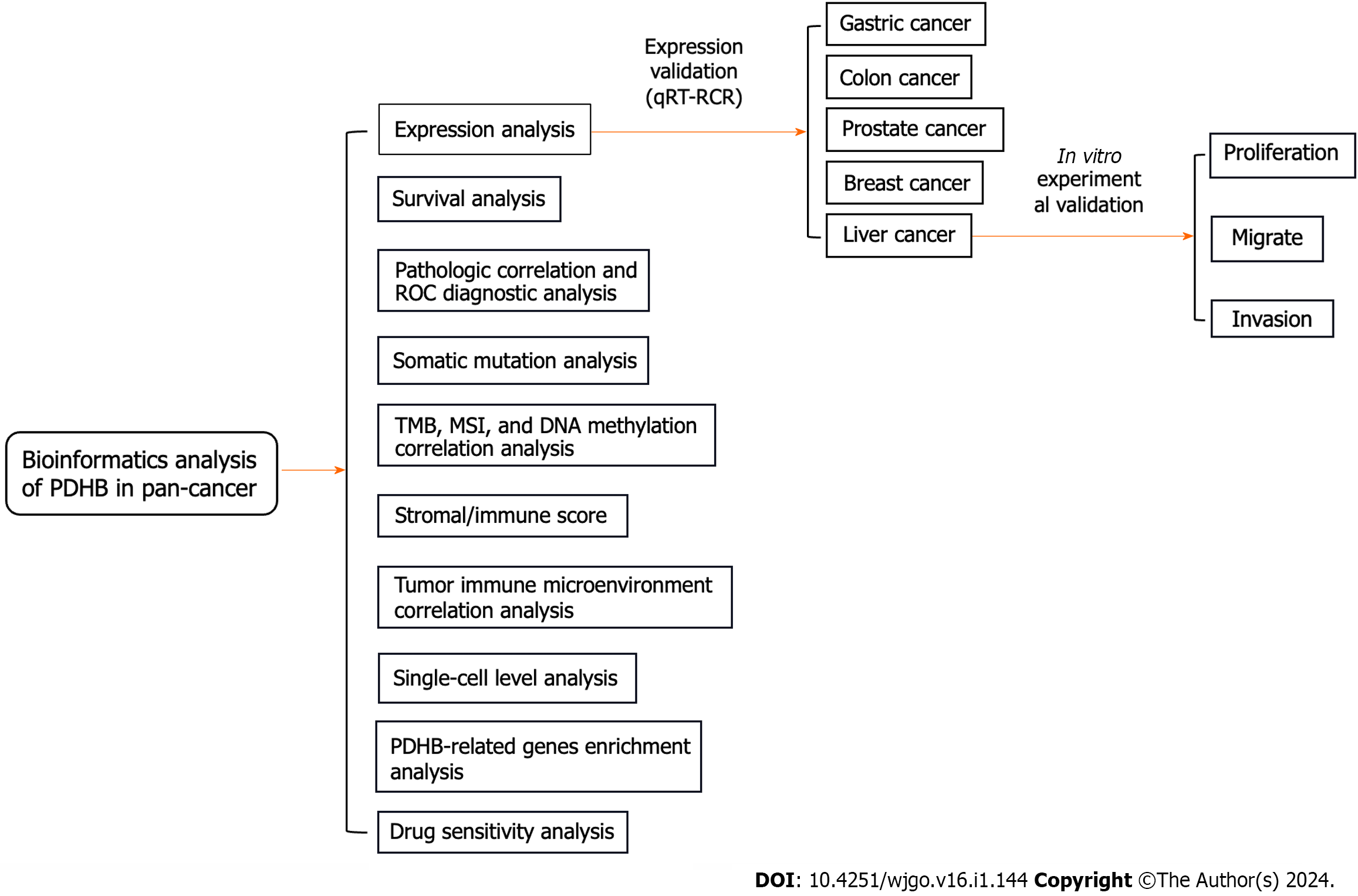
Figure 1 The predominant methodical workflow in the current study.
PDHB: Pyruvate dehydrogenase E1 subunit β; ROC: Receiver operating characteristic; TMB: Tumor mutation burden; MSI: Microsatellite instability; qRT-RCR: Real-time quantitative PCR.
Figure 2 Expression levels of pyruvate dehydrogenase E1 subunit β in pan-cancer.
A: Comparison of pyruvate dehydrogenase E1 subunit β (PDHB) expression levels in different cancers based on The Cancer Genome Atlas (TCGA) database; B: Expression of PDHB in cancer vs normal tissues in the TCGA database; C: Differential expression of PDHB in cancer and normal tissues in the TCGA joint Genotype Tissue Expression Dataset database; D: Protein levels of PDHB in tumors and normal tissues from the UALCAN database. aP < 0.05, bP < 0.01, cP < 0.001. PDHB: Pyruvate dehydrogenase E1 subunit β; ACC: Adrenocortical carcinoma; BLCA: Bladder urothelial carcinoma; BRCA: Breast invasive carcinoma; CESC: Cervical squamous cell carcinoma and Endocervical adenocarcinoma; CHOL: Cholangiocarcinoma; COAD: Colon adenocarcinoma; DLBC: Lymphoid neoplasm diffuse large B-cell lymphoma; ESCA: Esophageal carcinoma; GBM: Glioblastoma multiforme; HNSC: Head and Neck squamous cell carcinoma; KICH: Kidney chromophobe; KIRC: Kidney renal clear cell carcinoma; KIRP: Kidney renal papillary cell carcinoma; LAML: Acute myeloid leukemia; LGG: Brain lower grade glioma; LIHC: Liver hepatocellular carcinoma; LUAD: Lung adenocarcinoma; LUSC: Lung squamous cell carcinoma; MESO: Mesothelioma; OV: Ovarian serous cystadenocarcinoma; PAAD: Pancreatic adenocarcinoma; PCPG: Pheochromocytoma and Paraganglioma; PRAD: Prostate adenocarcinoma; READ: Rectum adenocarcinoma; SKCM: Skin cutaneous melanoma; STAD: Stomach adenocarcinoma; TGCT: Testicular germ cell tumors; THCA: Thyroid carcinoma; THYM: Thymoma; UCEC: Uterine corpus endometrial carcinoma; UCS: Uterine carcinosarcoma; UVM: Uveal melanoma.
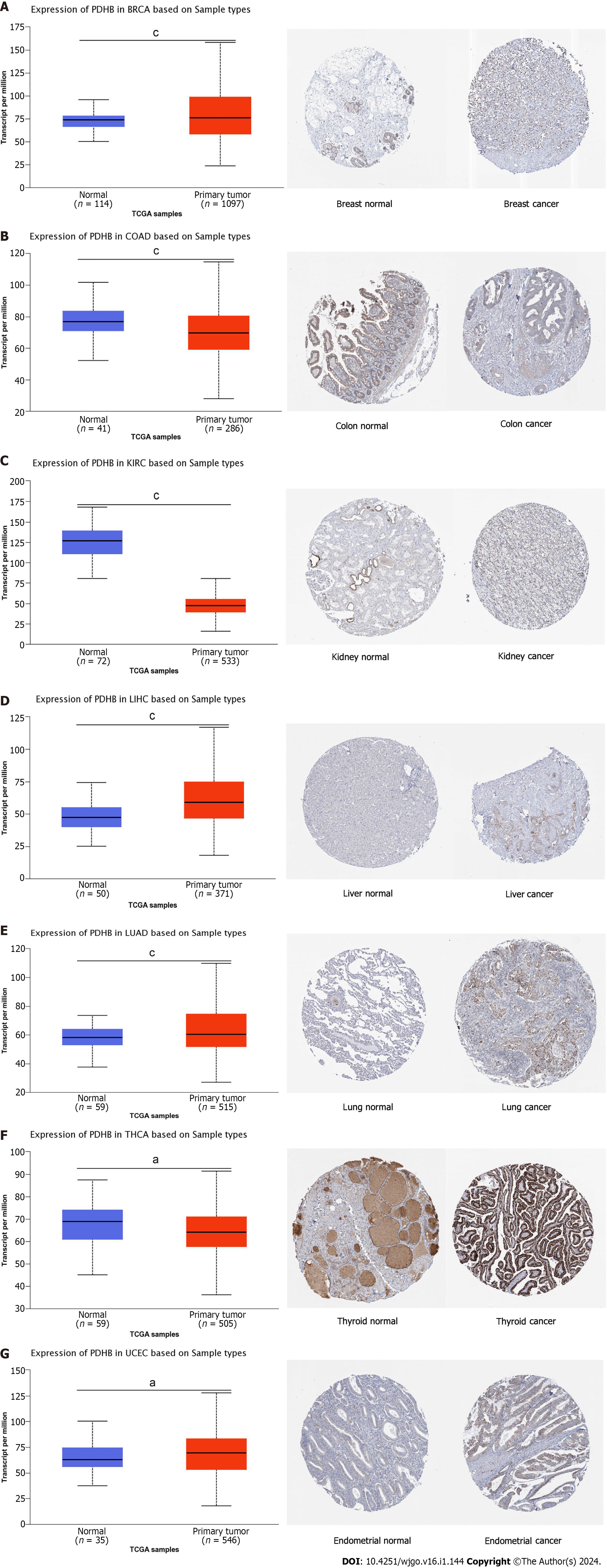
Figure 3 Pyruvate dehydrogenase E1 subunit β expression in the UACLAN and HPA databases.
A: Breast invasive carcinoma; B: Colon adenocarcinoma; C: Kidney renal clear cell carcinoma; D: Liver hepatocellular carcinoma; E: Lung adenocarcinoma; F: Thyroid carcinoma; G: Uterine corpus endometrial carcinoma. BRCA: Breast invasive carcinoma; COAD: Colon adenocarcinoma; KIRC: Kidney renal clear cell carcinoma; LIHC: Liver hepatocellular carcinoma; LUAD: Lung adenocarcinoma; THCA: Thyroid carcinoma; UCEC: Uterine corpus endometrial carcinoma.
Figure 4 The correlation analysis of pyruvate dehydrogenase E1 subunit β expression and prognosis in The Cancer Genome Atlas using COX regression analysis.
A: Overall survival; B: Progression-free survival; C: Disease-specific survival; D: Disease-free survival. OS: Overall survival; PFS: Progression-free survival; DSS: Disease-specific survival; DFS: Disease-free survival. ACC: Adrenocortical carcinoma; BLCA: Bladder urothelial carcinoma; BRCA: Breast invasive carcinoma; CESC: Cervical squamous cell carcinoma and Endocervical adenocarcinoma; CHOL: Cholangiocarcinoma; COAD: Colon adenocarcinoma; DLBC: Lymphoid neoplasm diffuse large B-cell lymphoma; ESCA: Esophageal carcinoma; GBM: Glioblastoma multiforme; HNSC: Head and Neck squamous cell carcinoma; KICH: Kidney chromophobe; KIRC: Kidney renal clear cell carcinoma; KIRP: Kidney renal papillary cell carcinoma; LAML: Acute myeloid leukemia; LGG: Brain lower grade glioma; LIHC: Liver hepatocellular carcinoma; LUAD: Lung adenocarcinoma; LUSC: Lung squamous cell carcinoma; MESO: Mesothelioma; OV: Ovarian serous cystadenocarcinoma; PAAD: Pancreatic adenocarcinoma; PCPG: Pheochromocytoma and Paraganglioma; PRAD: Prostate adenocarcinoma; READ: Rectum adenocarcinoma; SKCM: Skin cutaneous melanoma; STAD: Stomach adenocarcinoma; TGCT: Testicular germ cell tumors; THCA: Thyroid carcinoma; THYM: Thymoma; UCEC: Uterine corpus endometrial carcinoma; UCS: Uterine carcinosarcoma; UVM: Uveal melanoma.
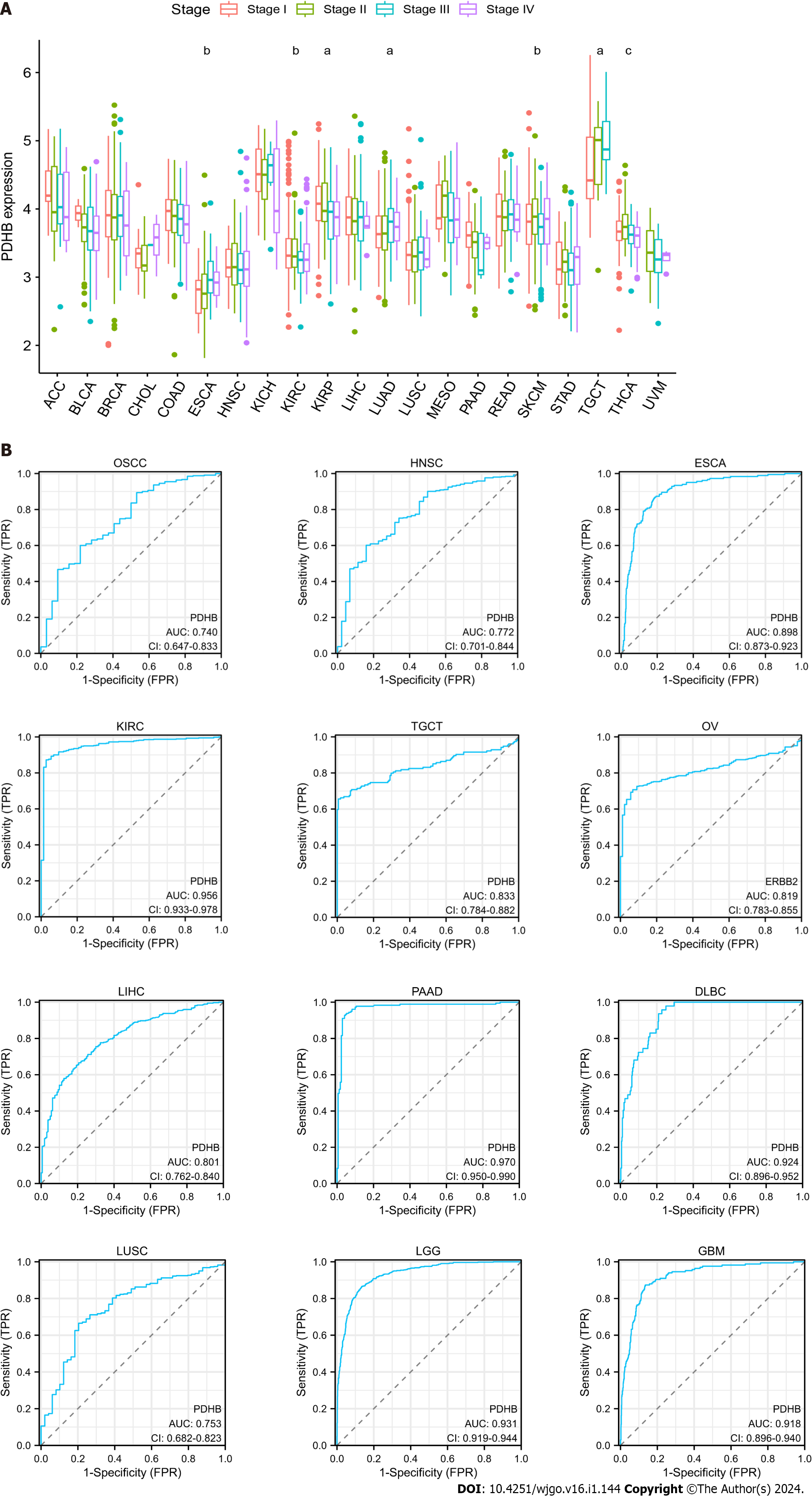
Figure 5 Correlation of pyruvate dehydrogenase E1 subunit β expression with tumor stage in multiple cancers and receiver operating characteristic diagnostic analysis.
A: Correlation between pyruvate dehydrogenase E1 subunit β (PDHB) gene expression and tumor staging; B: Receiver operating characteristic curve analysis of PDHB in various cancers (AreaUnderROC > 0.7). ACC: Adrenocortical carcinoma; BLCA: Bladder urothelial carcinoma; BRCA: Breast invasive carcinoma; CHOL: Cholangiocarcinoma; COAD: Colon adenocarcinoma; ESCA: Esophageal carcinoma; HNSC: Head and Neck squamous cell carcinoma; KICH: Kidney chromophobe; KIRC: Kidney renal clear cell carcinoma; KIRP: Kidney renal papillary cell carcinoma; LIHC: Liver hepatocellular carcinoma; LUAD: Lung adenocarcinoma; LUSC: Lung squamous cell carcinoma; MESO: Mesothelioma; PAAD: Pancreatic adenocarcinoma; PRAD: Prostate adenocarcinoma; SKCM: Skin cutaneous melanoma; STAD: Stomach adenocarcinoma; TGCT: Testicular germ cell tumors; THCA: Thyroid carcinoma; UVM: Uveal melanoma.
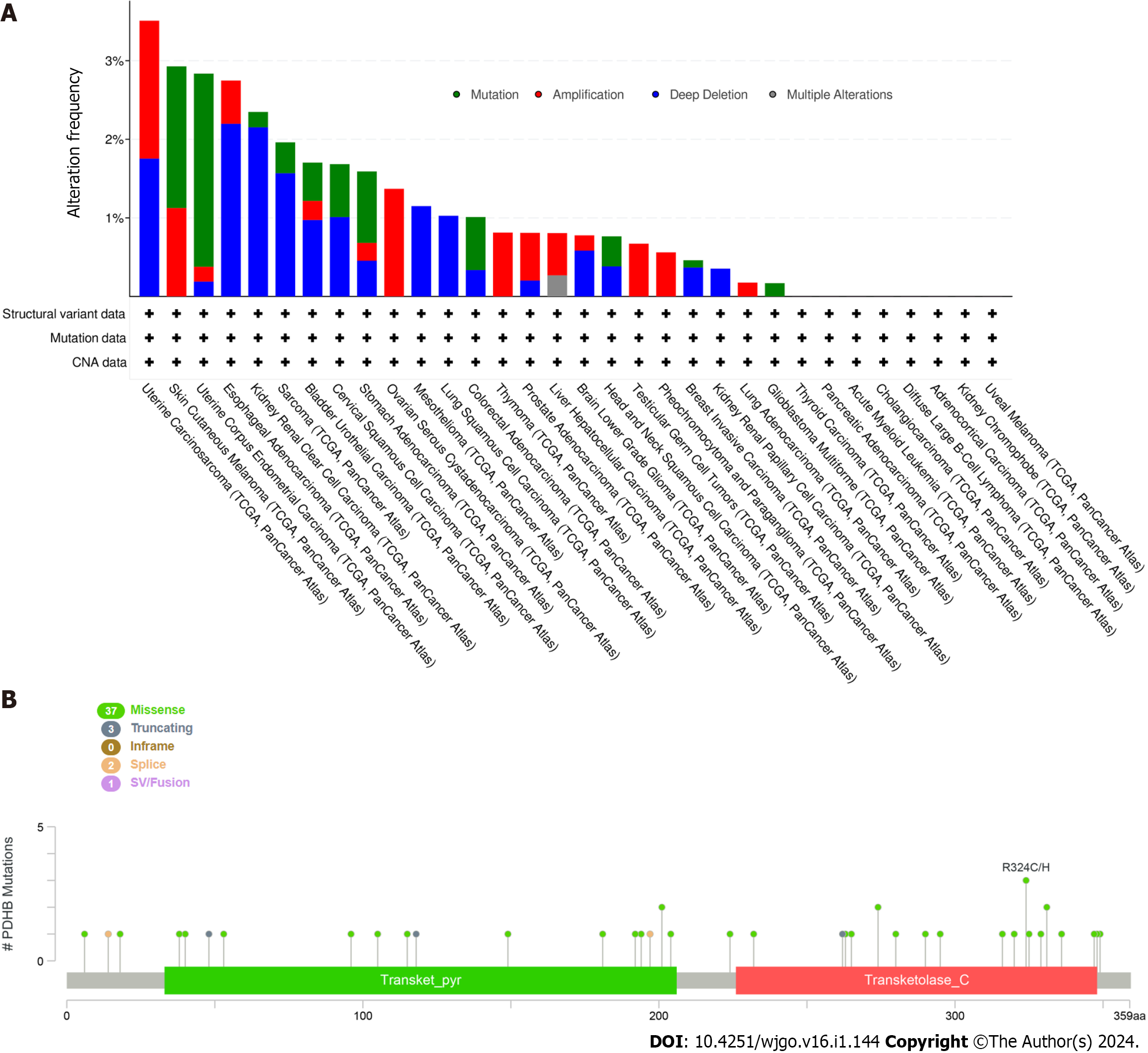
Figure 6 Mutation frequency of pyruvate dehydrogenase E1 subunit β in pan-cancer.
A: Somatic mutation analysis of pyruvate dehydrogenase E1 subunit β (PDHB) in different cancers; B: CBioPortal shows the mutation type and mutation frequency of PDHB sequences.
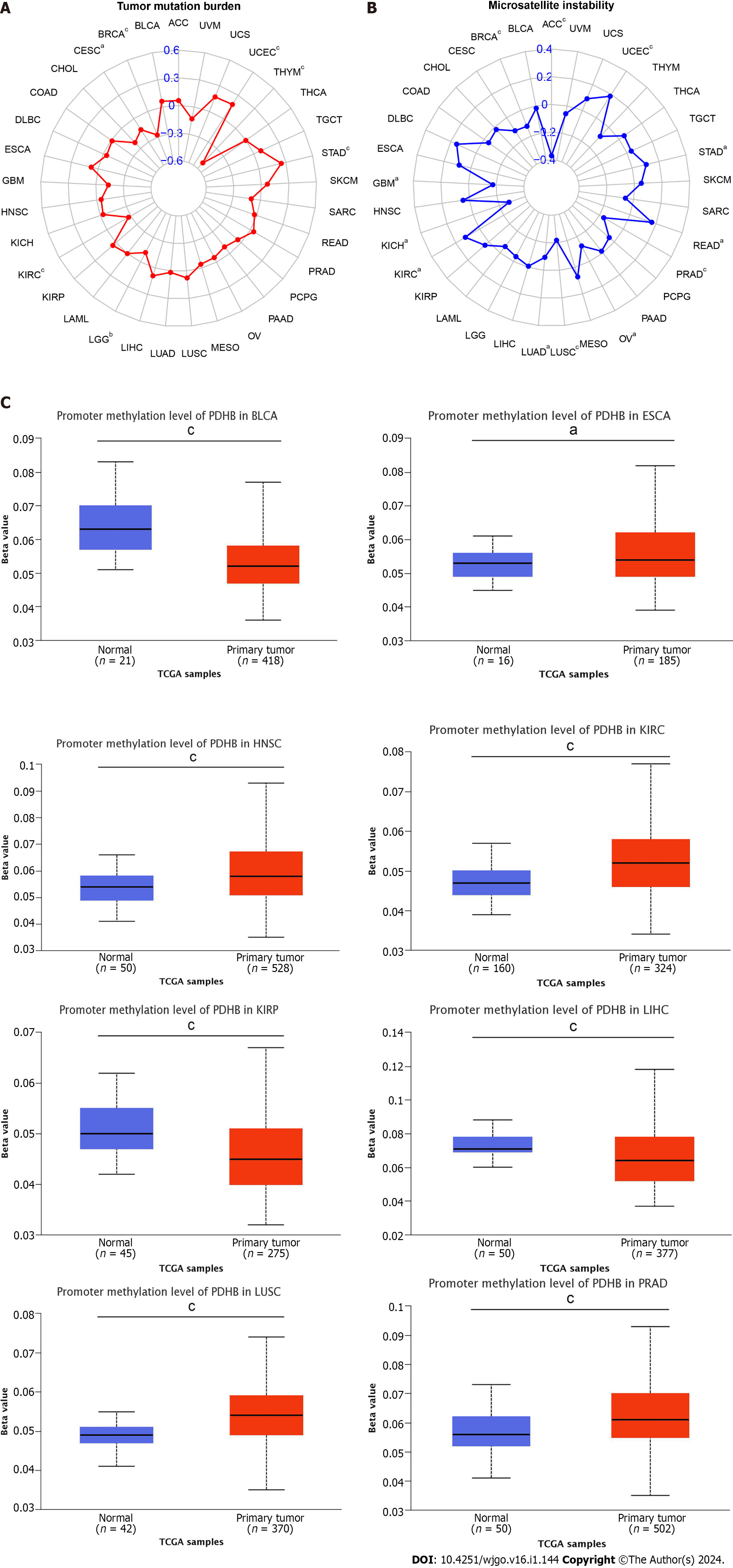
Figure 7 Correlation analysis of pyruvate dehydrogenase E1 subunit β gene expression with DNA methylation levels in tumor mutation burden, microsatellite instability and pan-cancer.
A: Radar plot demonstrating the relationship between tumor mutation burden and pyruvate dehydrogenase E1 subunit β (PDHB) gene expression in various malignancies. Correlation coefficients are indicated by red curves and ranges are indicated by blue values; B: Radar plot showing the relationship between microsatellite instability and PDHB gene expression and various malignancies. Correlation coefficients are shown by blue curves and ranges are shown by green values; C: PDHB methylation levels in the UALCAN database on different cancers. aP < 0.05, bP < 0.01, cP < 0.001. ACC: Adrenocortical carcinoma; BLCA: Bladder urothelial carcinoma; BRCA: Breast invasive carcinoma; CESC: Cervical squamous cell carcinoma and Endocervical adenocarcinoma; CHOL: Cholangiocarcinoma; COAD: Colon adenocarcinoma; DLBC: Lymphoid neoplasm diffuse large B-cell lymphoma; ESCA: Esophageal carcinoma; GBM: Glioblastoma multiforme; HNSC: Head and Neck squamous cell carcinoma; KICH: Kidney chromophobe; KIRC: Kidney renal clear cell carcinoma; KIRP: Kidney renal papillary cell carcinoma; LAML: Acute myeloid leukemia; LGG: Brain lower grade glioma; LIHC: Liver hepatocellular carcinoma; LUAD: Lung adenocarcinoma; LUSC: Lung squamous cell carcinoma; MESO: Mesothelioma; OV: Ovarian serous cystadenocarcinoma; PAAD: Pancreatic adenocarcinoma; PCPG: Pheochromocytoma and Paraganglioma; PRAD: Prostate adenocarcinoma; READ: Rectum adenocarcinoma; SKCM: Skin cutaneous melanoma; STAD: Stomach adenocarcinoma; TGCT: Testicular germ cell tumors; THCA: Thyroid carcinoma; THYM: Thymoma; UCEC: Uterine corpus endometrial carcinoma; UCS: Uterine carcinosarcoma; UVM: Uveal melanoma.
Figure 8 Correlation of pyruvate dehydrogenase E1 subunit β gene expression with stromal and immune scores in different cancers.
A: Correlation analysis of pyruvate dehydrogenase E1 subunit β (PDHB) gene expression with immune scores of bladder urothelial carcinoma (BLCA), breast invasive carcinoma (BRCA), brain lower grade glioma (LGG), lung adenocarcinoma (LUAD), lung squamous cell carcinoma, pancreatic adenocarcinoma (PAAD), thyroid carcinoma (THCA), uterine corpus endometrial carcinoma (UCEC); B: Correlation analysis of PDHB gene expression with stromal scores of BLCA, BRCA, LGG, liver hepatocellular carcinoma, LUAD, mesothelioma, ovarian serous cystadenocarcinoma, PAAD, prostate adenocarcinoma, sarcoma, testicular germ cell tumors, THCA, thymoma, UCEC. aP < 0.05, bP < 0.01, cP < 0.001. BRCA: Breast invasive carcinoma; BLCA: Bladder urothelial carcinoma; LGG: Brain lower grade glioma; LUAD: Lung adenocarcinoma; LUSC: Lung squamous cell carcinoma; PAAD: Pancreatic adenocarcinoma; THCA: Thyroid carcinoma; UCEC: Uterine corpus endometrial carcinoma; BRCA: Breast invasive carcinoma; LIHC: Liver hepatocellular carcinoma; MESO: Mesothelioma; OV: Ovarian serous cystadenocarcinoma; PRAD: Prostate adenocarcinoma; SARC: Sarcoma; TGCT: Testicular germ cell tumors; THCA: Thyroid carcinoma; THYM: Thymoma; UCEC: Uterine corpus endometrial carcinoma.
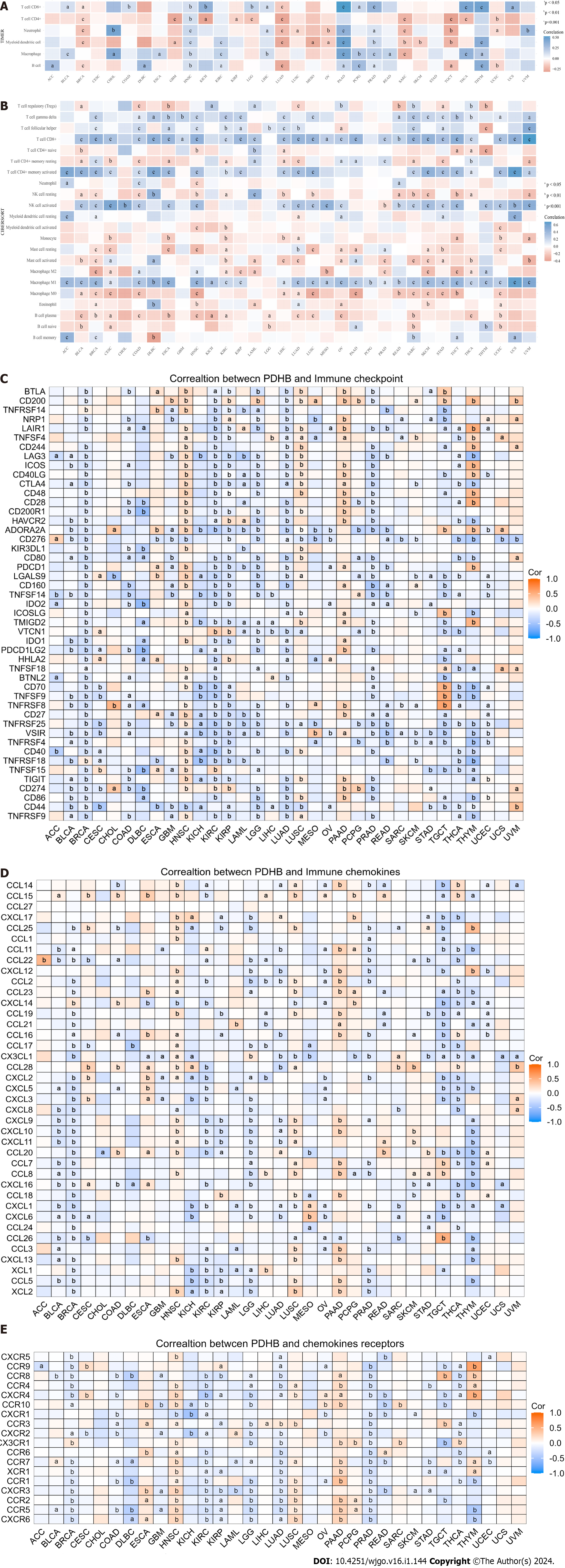
Figure 9 Correlation analysis of pyruvate dehydrogenase E1 subunit β expression and tumor immunity.
A: TIMER method to analyze the correlation between pyruvate dehydrogenase E1 subunit β (PDHB) and immune cell infiltration; B: CIBERSORT method to analyze PDHB correlation with immune cell infiltration; C: Co-expression analysis of PDHB with tumor chemokines; D: Co-expression analysis of PDHB with tumor chemokine receptors; E: Co-expression analysis of PDHB with immune checkpoint gene. aP < 0.05, bP < 0.01, cP < 0.001. PDHB: Pyruvate dehydrogenase E1 subunit β. ACC: Adrenocortical carcinoma; BLCA: Bladder urothelial carcinoma; BRCA: Breast invasive carcinoma; CESC: Cervical squamous cell carcinoma and Endocervical adenocarcinoma; CHOL: Cholangiocarcinoma; COAD: Colon adenocarcinoma; DLBC: Lymphoid neoplasm diffuse large B-cell lymphoma; ESCA: Esophageal carcinoma; GBM: Glioblastoma multiforme; HNSC: Head and Neck squamous cell carcinoma; KICH: Kidney chromophobe; KIRC: Kidney renal clear cell carcinoma; KIRP: Kidney renal papillary cell carcinoma; LAML: Acute myeloid leukemia; LGG: Brain lower grade glioma; LIHC: Liver hepatocellular carcinoma; LUAD: Lung adenocarcinoma; LUSC: Lung squamous cell carcinoma; MESO: Mesothelioma; OV: Ovarian serous cystadenocarcinoma; PAAD: Pancreatic adenocarcinoma; PCPG: Pheochromocytoma and Paraganglioma; PRAD: Prostate adenocarcinoma; READ: Rectum adenocarcinoma; SKCM: Skin cutaneous melanoma; STAD: Stomach adenocarcinoma; TGCT: Testicular germ cell tumors; THCA: Thyroid carcinoma; THYM: Thymoma; UCEC: Uterine corpus endometrial carcinoma; UCS: Uterine carcinosarcoma; UVM: Uveal melanoma.
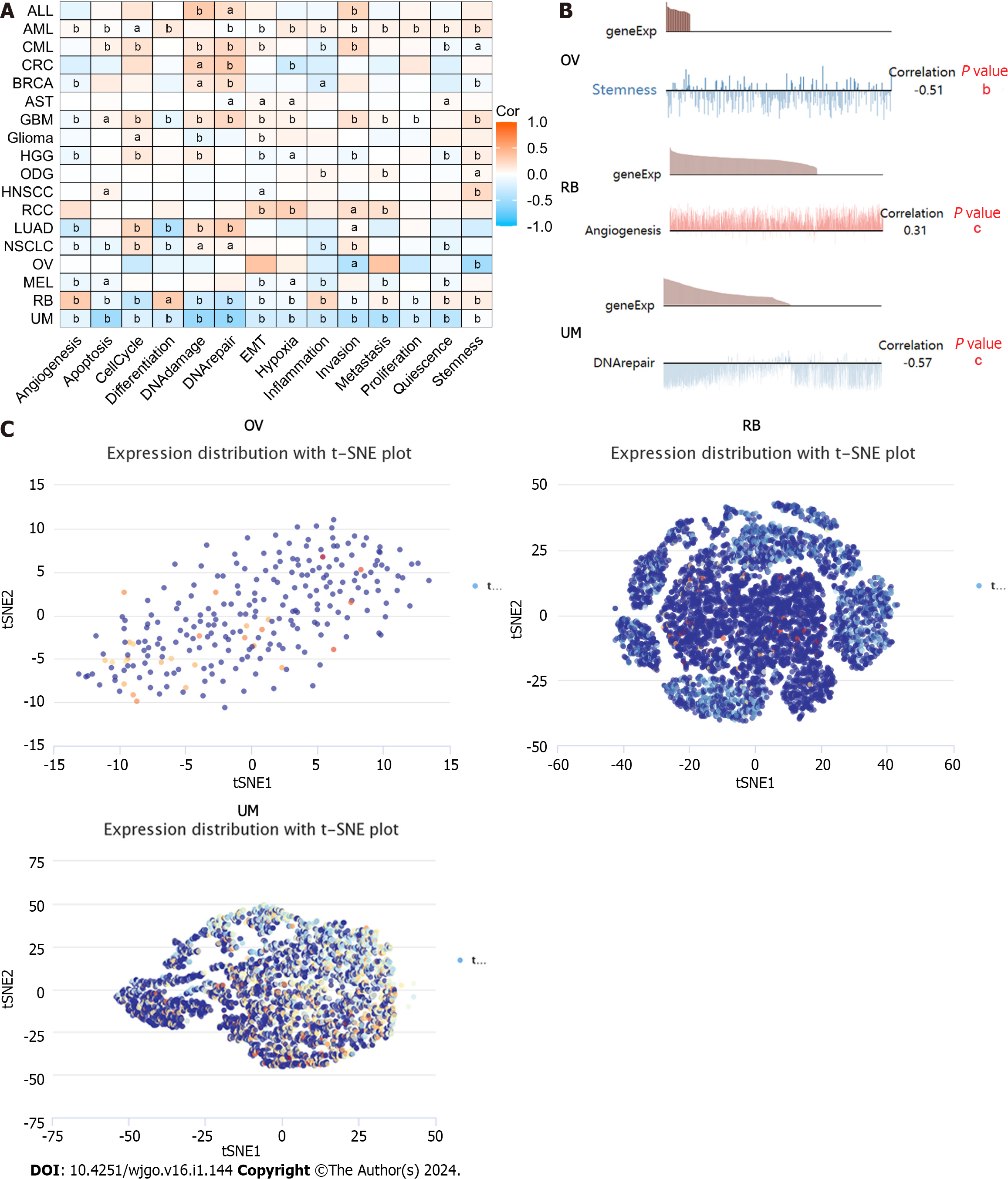
Figure 10 Pyruvate dehydrogenase E1 subunit β expression levels at the single-cell sequencing level.
A and B: CancerSEA database demonstrates the correlation of pyruvate dehydrogenase E1 subunit β (PDHB) expression with multiple biological functions in pan-cancer; C: t-Distributed Stochastic Neighbor Embedding plots showing the distribution of PDHB in ovarian serous cystadenocarcinoma, retinoblastoma, and uveal melanoma at the single-cell level. aP < 0.05, bP < 0.01, cP < 0.001. OV: Ovarian serous cystadenocarcinoma; RB: Retinoblastoma; UM: Uveal melanoma.
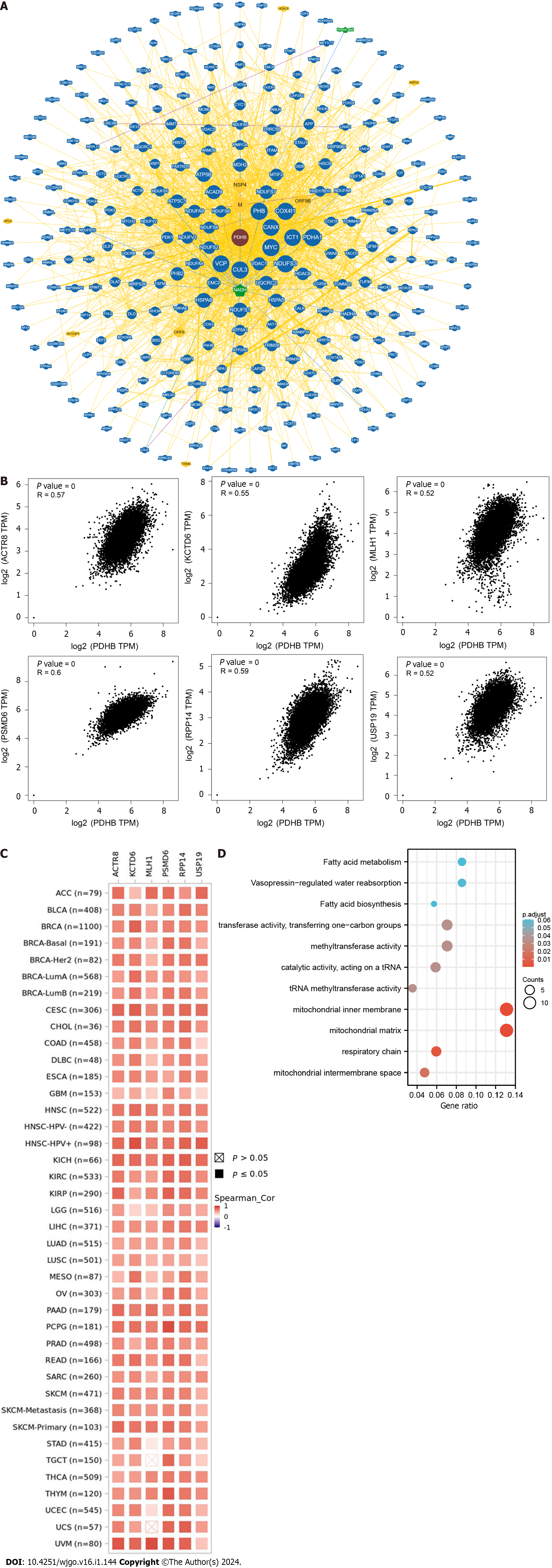
Figure 11 Enrichment analysis of pyruvate dehydrogenase E1 subunit β-associated genes in pan-cancer.
A: Interaction network of pyruvate dehydrogenase E1 subunit β (PDHB)-associated biomarkers derived from the BioGRID database; B: GEPIA2.0 showed that PDHB expression was positively correlated with actin related protein 8 (ACTR8), potassium channel tetramerization domain containing 6 (KCTD6), mutl homolog 1 (MLH1), proteasome 26s subunit, non-ATPase 6 (PSMD6), ribonuclease P/MRP subunit p14 (RPP14), and ubiquitin specific peptidase 19 (USP19) genes; C: Heat map showing PDHB expression positively correlated with 6 genes (ACTR8, KCTD6, MLH1, PSMD6, RPP14, USP19); D: Enrichment analysis of PDHB-related genes. PDHB: Pyruvate dehydrogenase E1 subunit β. ACC: Adrenocortical carcinoma; BLCA: Bladder urothelial carcinoma; BRCA: Breast invasive carcinoma; CESC: Cervical squamous cell carcinoma and Endocervical adenocarcinoma; CHOL: Cholangiocarcinoma; COAD: Colon adenocarcinoma; DLBC: Lymphoid neoplasm diffuse large B-cell lymphoma; ESCA: Esophageal carcinoma; GBM: Glioblastoma multiforme; HNSC: Head and Neck squamous cell carcinoma; KICH: Kidney chromophobe; KIRC: Kidney renal clear cell carcinoma; KIRP: Kidney renal papillary cell carcinoma; LAML: Acute myeloid leukemia; LGG: Brain lower grade glioma; LIHC: Liver hepatocellular carcinoma; LUAD: Lung adenocarcinoma; LUSC: Lung squamous cell carcinoma; MESO: Mesothelioma; OV: Ovarian serous cystadenocarcinoma; PAAD: Pancreatic adenocarcinoma; PCPG: Pheochromocytoma and Paraganglioma; PRAD: Prostate adenocarcinoma; READ: Rectum adenocarcinoma; SKCM: Skin cutaneous melanoma; STAD: Stomach adenocarcinoma; TGCT: Testicular germ cell tumors; THCA: Thyroid carcinoma; THYM: Thymoma; UCEC: Uterine corpus endometrial carcinoma; UCS: Uterine carcinosarcoma; UVM: Uveal melanoma.
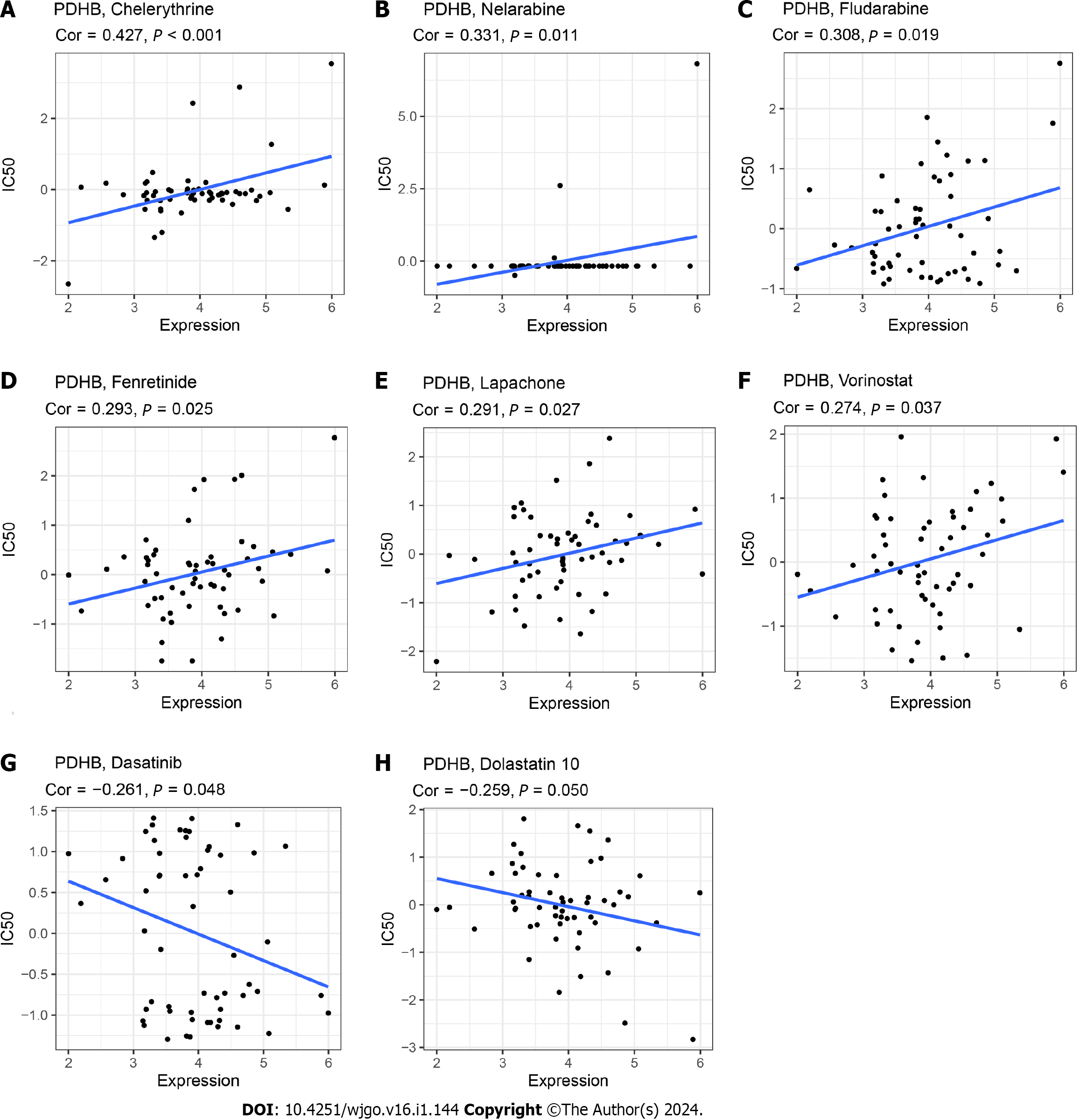
Figure 12 Drug sensitivity analysis.
Correlation of IC50 with pyruvate dehydrogenase E1 subunit β expression for different drugs. A: Chelerythrine; B: Nelarabine; C: Fludarabine; D: Fenretinide; E: Lapachone; F: Vorinostat; G: Dasatinib; H: Dolastatin 10. PDHB: Pyruvate dehydrogenase E1 subunit β.
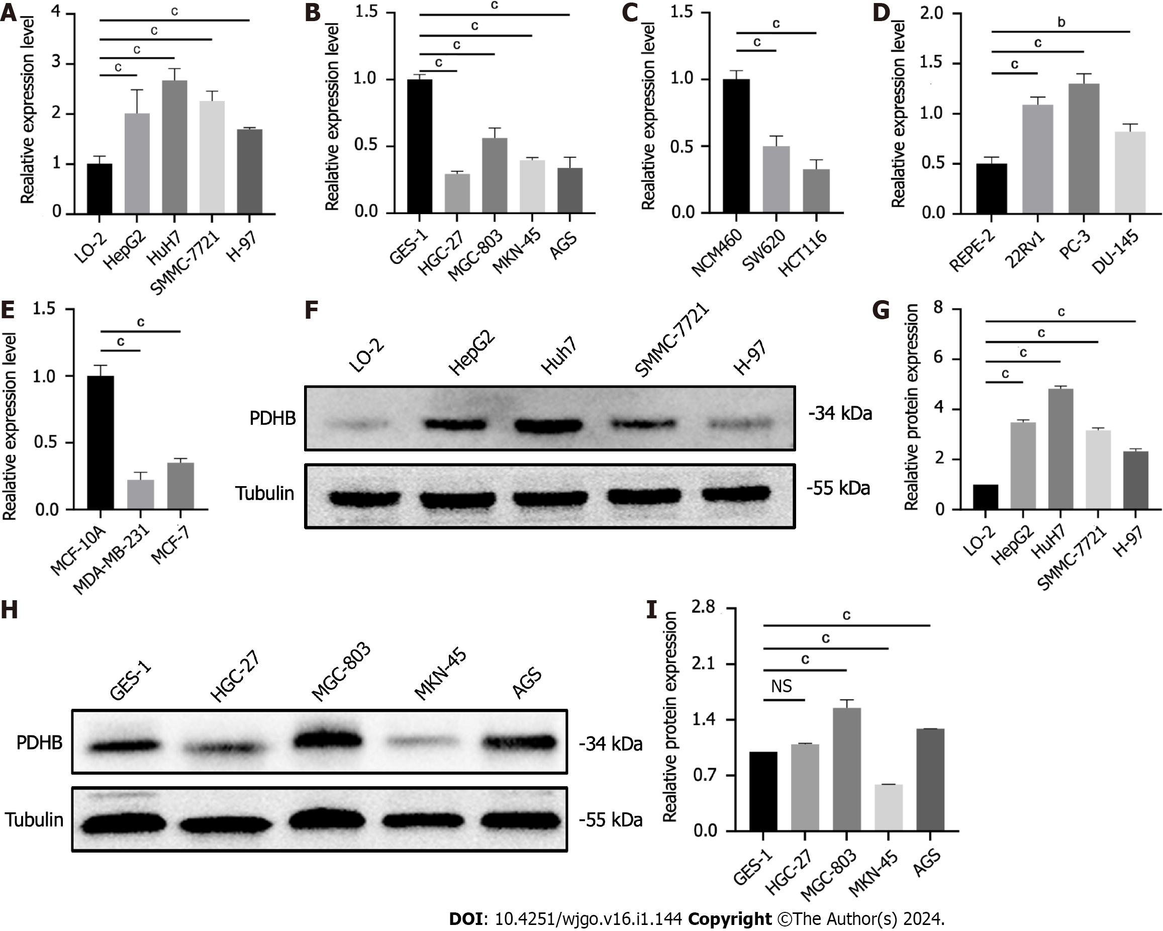
Figure 13 Results of pyruvate dehydrogenase E1 subunit β expression validation.
A: Pyruvate dehydrogenase E1 subunit β (PDHB) expression in the human normal hepatocyte cell line (L-O2) and human hepatoma cell lines (SMMC-7721, HepG2, Huh7, H-97); B: PDHB expression in the human normal gastric mucosal cell line (GES-1) and human gastric cancer cell lines (HGC-27, MGC-803, MKN-45); C: PDHB expression in the human normal colonic epithelial cell line (NCM460) and human colon cancer cell lines (SW620, HCT116); D: PDHB expression in the human normal breast cell line (MCF-10A) and breast cancer cell lines (MDA-MB-231, MCF-7); E: PDHB expression in the human normal prostate cell line (RWPE-2) and prostate cancer cell lines (PC-3, 22Rv1 and DU145); F: Validation of PDHB protein expression in the L-O2 and hepatoma cell lines (HepG2, SMMC-7721, Huh7, H-97); G: Quantitative plots; H: Validation of PDHB protein expression in the GES-1 and gastric cancer cell lines (MKN-45, AGS, HGC-27, MGC-803); I: Quantitative plots. aP < 0.05, bP < 0.01, cP < 0.001. PDHB: Pyruvate dehydrogenase E1 subunit β; L-O2: Human normal hepatocyte cell line; GES-1: Gastric mucosal cells; NS: Not significant.
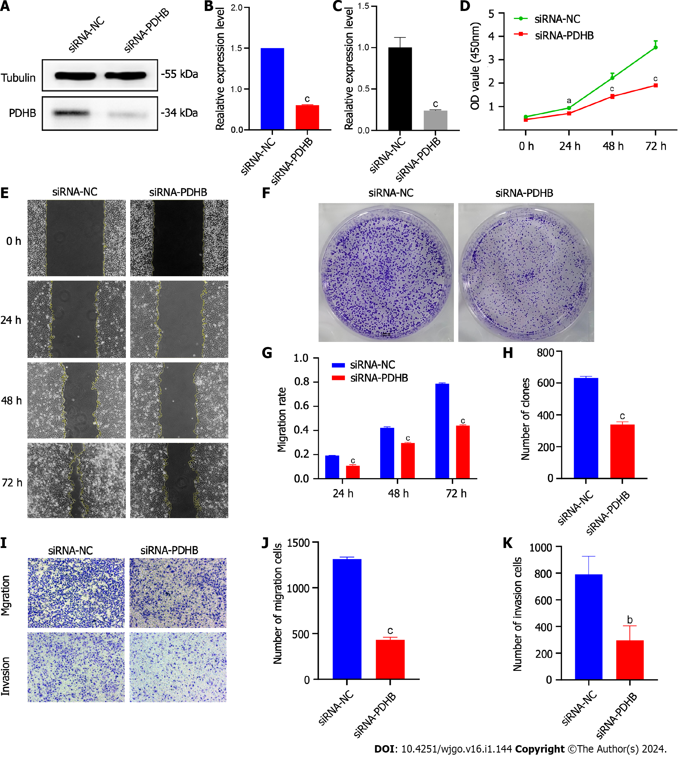
Figure 14 Effects of siRNA-pyruvate dehydrogenase E1 subunit β on proliferation, migration and invasion of Huh7 hepatoma cells.
A: Transfection efficiency of siRNA-pyruvate dehydrogenase E1 subunit β (PDHB) in Huh7 cell lines; B: The relative expression of mRNA reflects the efficiency of siRNA-PDHB transfection; C: siRNA-PDHB transfection efficiency by protein expression level; D: The CCK-8 method detected the proliferation capacity of Huh7 cell lines; E: Colony formation assay to determine the proliferation capacity of Huh7 cell lines; F: The histogram shows the number of colonies formed; G: Cell scratch assay to detect the migration capacity of Huh7 cell lines; H: Histogram of quantification of cell scratch assay results; I: Transwell method was used to detect the migration and invasion capacity of Huh7 cell lines; J: The histogram shows the number of migrating cells; K: The histogram shows the number of invading cells. aP < 0.05, bP < 0.01, cP < 0.001. PDHB: Pyruvate dehydrogenase E1 subunit β.
- Citation: Rong Y, Liu SH, Tang MZ, Wu ZH, Ma GR, Li XF, Cai H. Analysis of the potential biological value of pyruvate dehydrogenase E1 subunit β in human cancer. World J Gastrointest Oncol 2024; 16(1): 144-181
- URL: https://www.wjgnet.com/1948-5204/full/v16/i1/144.htm
- DOI: https://dx.doi.org/10.4251/wjgo.v16.i1.144


