Copyright
©The Author(s) 2023.
World J Gastrointest Oncol. Sep 15, 2023; 15(9): 1531-1543
Published online Sep 15, 2023. doi: 10.4251/wjgo.v15.i9.1531
Published online Sep 15, 2023. doi: 10.4251/wjgo.v15.i9.1531
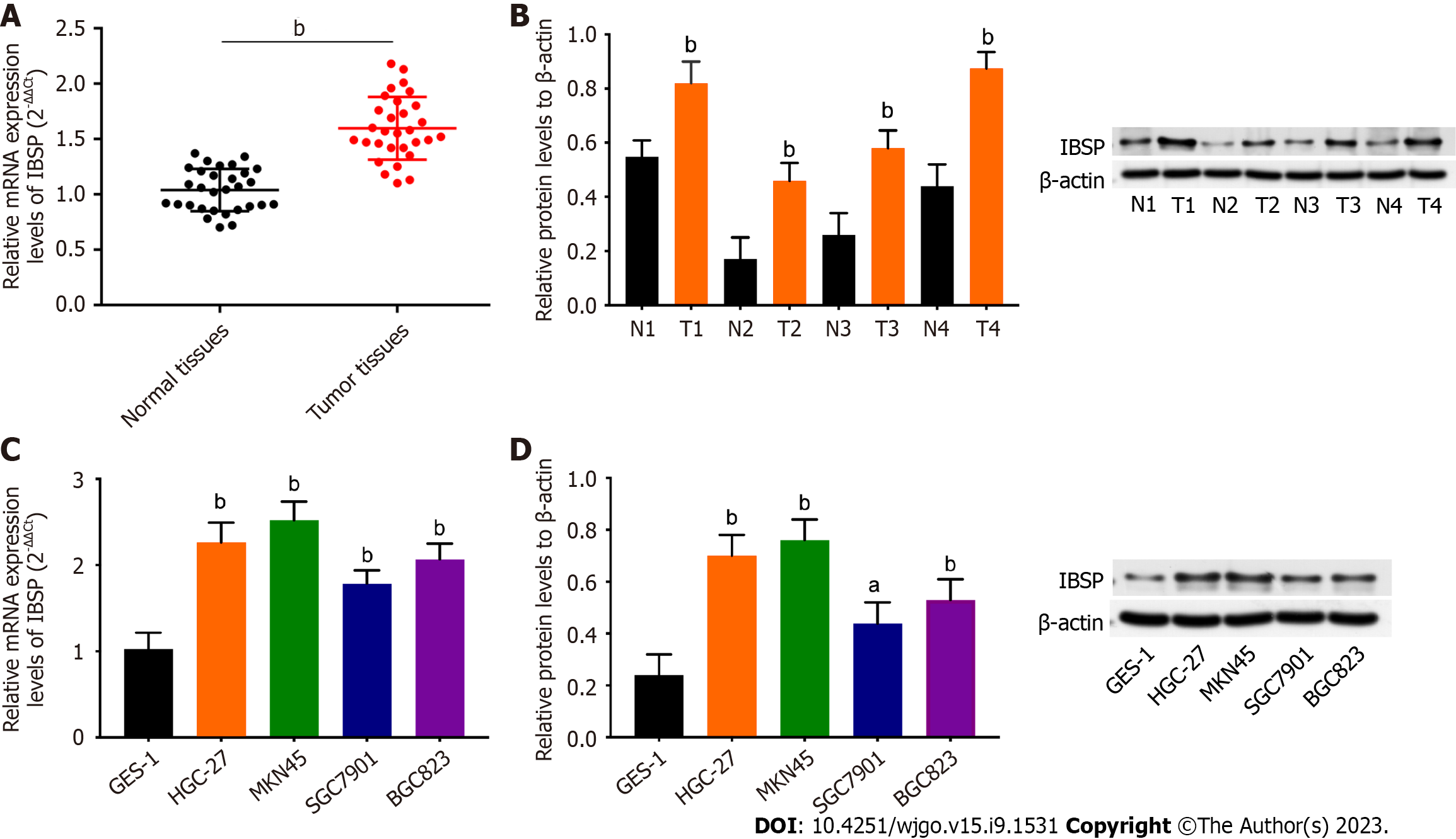
Figure 1 Integrin binding sialoprotein shows higher expression in gastric cancer tissues and cell lines.
A: The mRNA expression of integrin binding sialoprotein (IBSP) in gastric cancer (GC) tissues and normal adjacent tissues was examined by real-time quantitative polymerase chain reaction (RT-qPCR); B: The protein expression of IBSP in four pairs of GC tissues and normal adjacent tissues was detected by Western blot; C and D: The mRNA and protein expression levels of IBSP in gastric epithelial cell line (GES-1) and GC cell lines (HGC-27, MKN45, SGC-7901, and BGC823) were detected by RT-qPCR and Western blot, respectively. aP < 0.05, bP < 0.01. IBSP: Integrin binding sialoprotein.
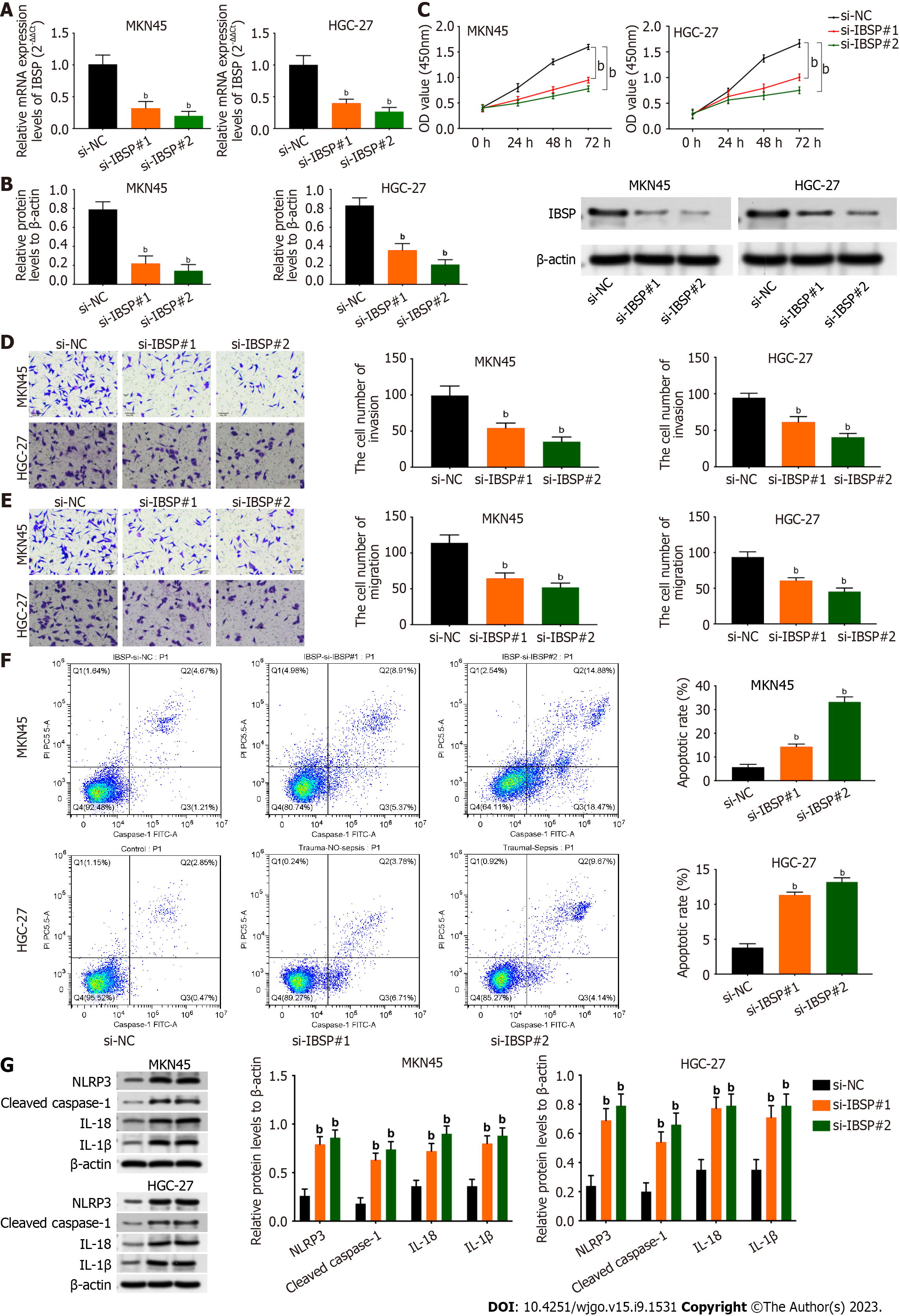
Figure 2 Integrin binding sialoprotein downregulation suppresses cell proliferation, migration, and invasion and facilitates pyroptosis.
A and B: The mRNA and protein expression levels of integrin binding sialoprotein (IBSP) were assessed in the si-NC, si-IBSP#1, and si-IBSP#2 groups by real-time quantitative polymerase chain reaction and Western blot in MKN45 and HGC-27 cells, respectively; C: Cell viability was detected after silencing IBSP by Cell-Counting Kit-8 assay in MKN45 and HGC-27 cells; D and E: Cell invasion and migration were evaluated by Transwell assay in MKN45 and HGC-27 cells; F: Pyroptosis was measured after IBSP knockdown by flow cytometry in MKN45 and HGC-27 cells; G: The protein expression levels of NLR family pyrin domain containing 3, cleaved caspase-1, interleukin-18 (IL-18), and IL-1β were examined by Western blot after suppressing IBSP in MKN45 and HGC-27 cells. bP < 0.01. IBSP: Integrin binding sialoprotein; IL: Interleukin; NLRP3: NLR family pyrin domain containing 3.
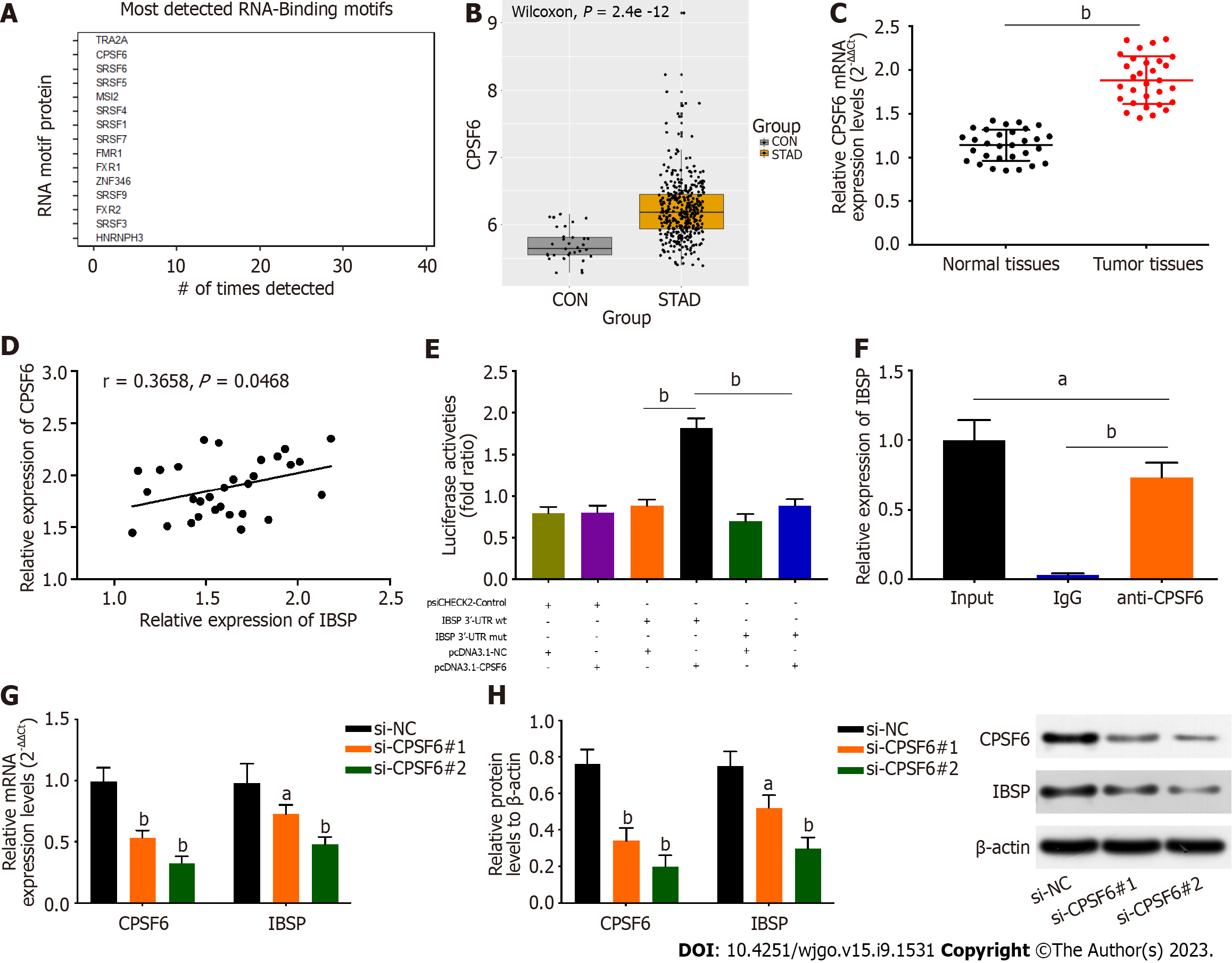
Figure 3 The RNA-binding protein cleavage and polyadenylation factor 6 binds to the 3’-untranslated region of integrin binding sialoprotein and regulates its expression.
A: Potential RNA-binding proteins that can bind to integrin binding sialoprotein (IBSP) were analyzed through bioinformatic analysis; B: The levels of cleavage and polyadenylation factor 6 (CPSF6) in stomach adenocarcinoma; C: The mRNA expression of CPSF6 was detected in GC tissues and normal adjacent tissues by real-time quantitative polymerase chain reaction (RT-qPCR); D: The correlation between CPSF6 and IBSP was verified; E and F: The binding ability between CPSF6 and IBSP was confirmed by luciferase reporter and RNA immunoprecipitation chip assays; G and H: The mRNA and protein expression of CPSF6 and IBSP was measured in the si-NC, si-CPSF6#1, and si-CPSF6#2 groups by RT-qPCR and Western blot, respectively. aP < 0.05, bP < 0.01.
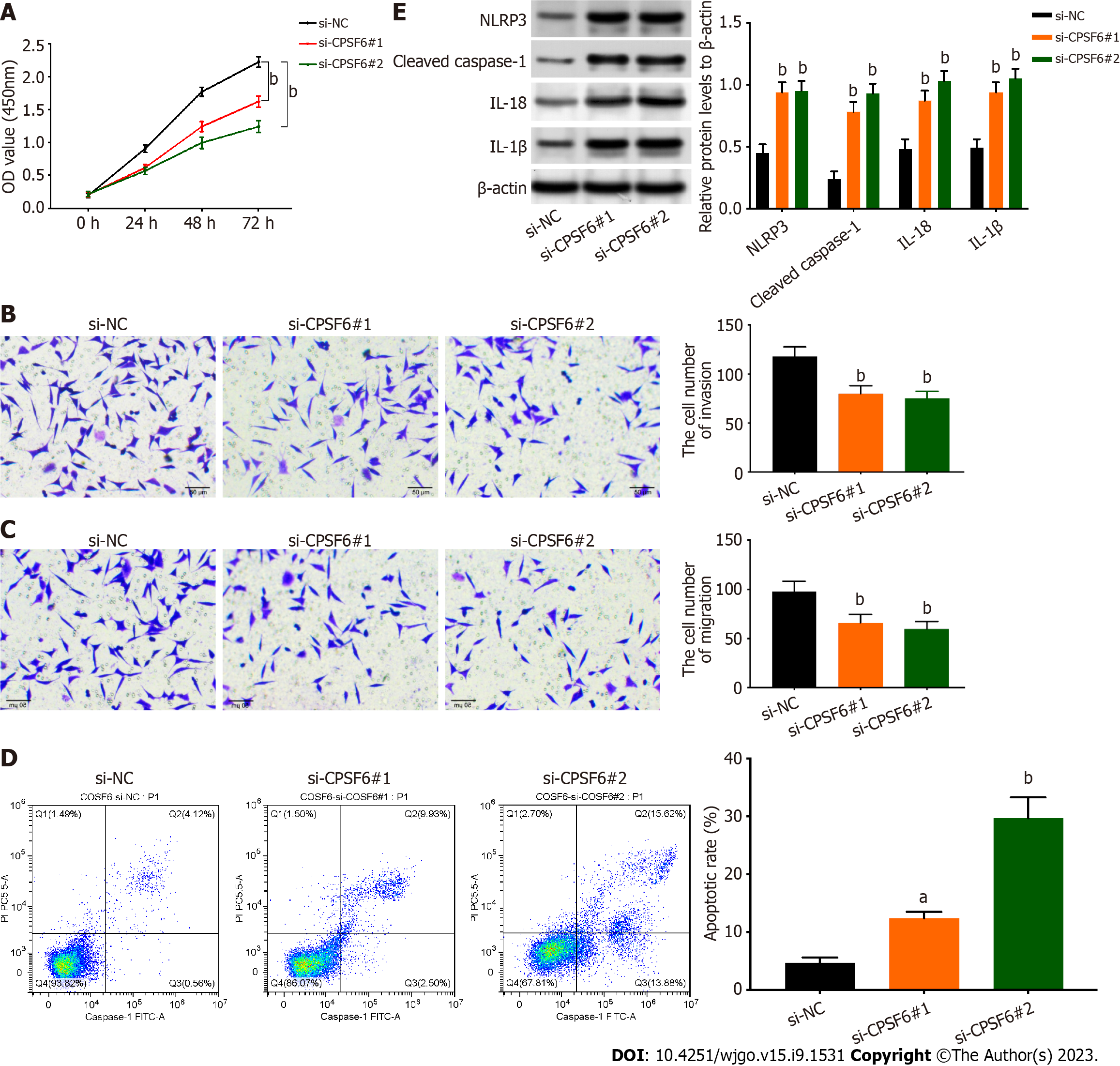
Figure 4 Knockdown of cleavage and polyadenylation factor 6 inhibits cell proliferation, migration, and invasion but boosts pyroptosis.
A: Cell viability was verified after suppressing cleavage and polyadenylation factor 6 (CPSF6) by Cell Counting Kit-8 assay; B and C: Cell migration and invasion were detected after inhibiting CPSF6 by Transwell assay; D: Cell apoptosis was examined after silencing CPSF6 by flow cytometry; E: The protein expression levels of NLR family pyrin domain containing 3, cleaved caspase-1, interleukin-18 (IL-18), and IL-1β were examined after suppressing CPSF6 by Western blot. aP < 0.05, bP < 0.01. CPSF6: Cleavage and polyadenylation factor 6; NLRP3: NLR family pyrin domain containing 3; IL: Interleukin.
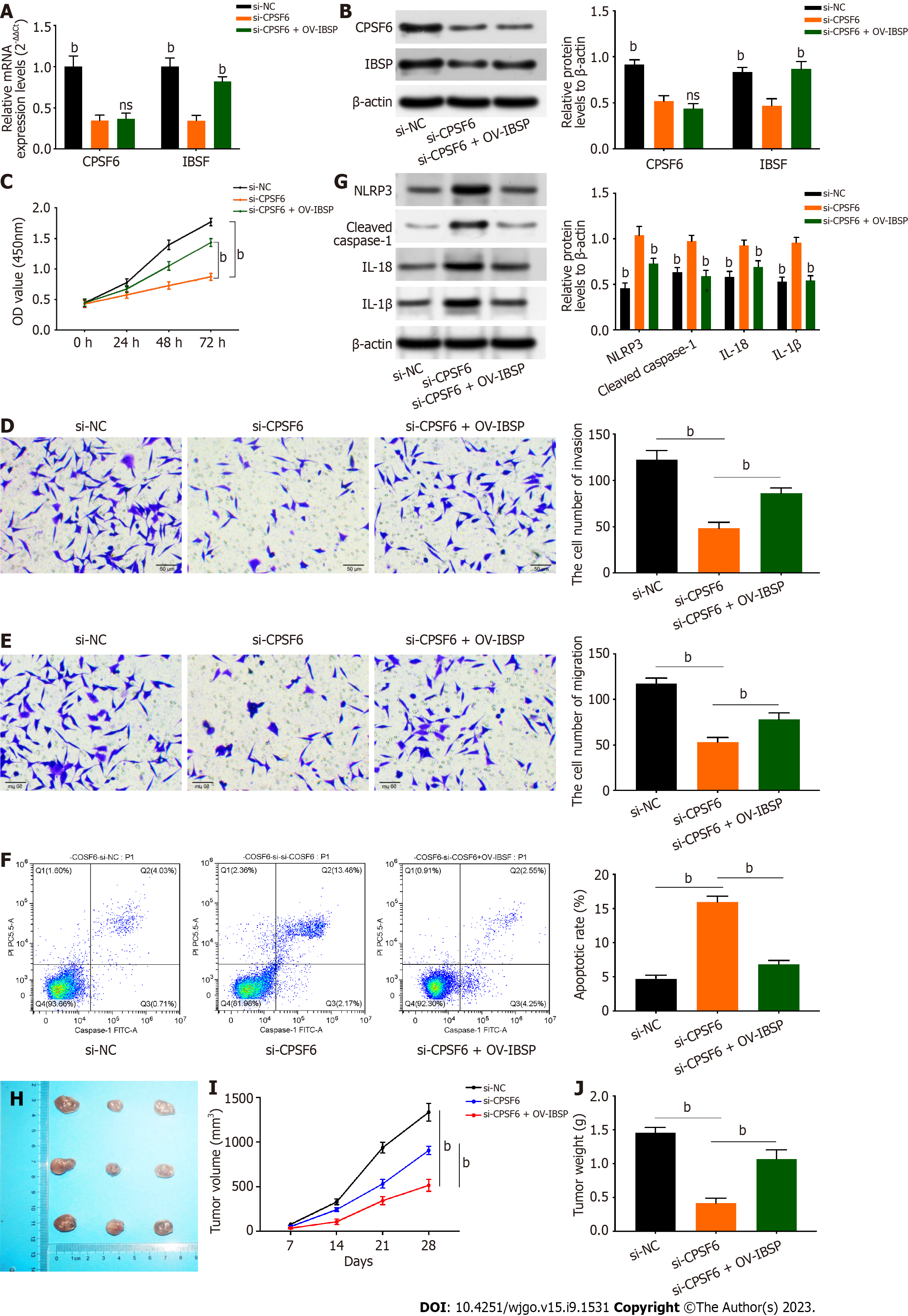
Figure 5 Cleavage and polyadenylation factor 6 regulates integrin binding sialoprotein to affect gastric cancer progression.
Cells were divided into the si-NC, si-cleavage and polyadenylation factor 6 (si-CPSF6), and si-CPSF6 + OV-integrin binding sialoprotein (IBSP) groups. A and B: The mRNA and protein expression of CPSF6 and IBSP was detected by real-time quantitative polymerase chain reaction (RT-qPCR) and Western blot, respectively; C: Cell viability was examined by Cell Counting Kit-8 assay; D and E: Cell invasion and migration were tested by transwell assay; F: Cell apoptosis was measured by flow cytometry; G: The protein expression levels of NLR family pyrin domain containing 3, cleaved caspase-1, interleukin-18 (IL-18), and IL-1β were detected by Western blot; H-J: Tumor size, volume, and weight were measured. bP < 0.01. IBSP: Integrin binding sialoprotein; CPSF6: Cleavage and polyadenylation factor 6; NLRP3: NLR family pyrin domain containing 3; IL: Interleukin.
- Citation: Wang XJ, Liu Y, Ke B, Zhang L, Liang H. RNA-binding protein CPSF6 regulates IBSP to affect pyroptosis in gastric cancer. World J Gastrointest Oncol 2023; 15(9): 1531-1543
- URL: https://www.wjgnet.com/1948-5204/full/v15/i9/1531.htm
- DOI: https://dx.doi.org/10.4251/wjgo.v15.i9.1531


