Copyright
©The Author(s) 2023.
World J Gastrointest Oncol. Jun 15, 2023; 15(6): 1096-1104
Published online Jun 15, 2023. doi: 10.4251/wjgo.v15.i6.1096
Published online Jun 15, 2023. doi: 10.4251/wjgo.v15.i6.1096
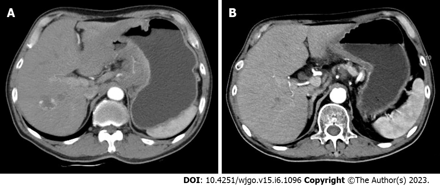
Figure 1 Enhanced computed tomography images prior to and after combined treatment.
A: Enhanced computed tomography (CT) before any treatment showed a lesion in the gastric wall; B: Enhanced CT after combined treatment revealed that the lesion was apparently decreased in size.
Figure 2 Fluorodeoxyglucose positron emission tomography/computed tomography images prior to and after combined treatment.
A: Fluorodeoxyglucose positron emission tomography/computed tomography (FDG PET/CT) prior to any treatment showed a large gastric mass with hypermetabolism, and the lesion was clearly decreased in size and metabolism after combined treatment; B: FDG PET/CT prior to any treatment showed hypermetabolism in the left supraclavicular area, and the lesion was clearly decreased in size and metabolism after combined treatment; C: FDG PET/CT prior to any treatment showed hypermetabolism in the left acetabular area, and the lesion was clearly decreased in size and metabolism after combined treatment.
- Citation: Zhou ML, Xu RN, Tan C, Zhang Z, Wan JF. Advanced gastric cancer achieving major pathologic regression after chemoimmunotherapy combined with hypofractionated radiotherapy: A case report. World J Gastrointest Oncol 2023; 15(6): 1096-1104
- URL: https://www.wjgnet.com/1948-5204/full/v15/i6/1096.htm
- DOI: https://dx.doi.org/10.4251/wjgo.v15.i6.1096


