Copyright
©The Author(s) 2023.
World J Gastrointest Oncol. Mar 15, 2023; 15(3): 562-570
Published online Mar 15, 2023. doi: 10.4251/wjgo.v15.i3.562
Published online Mar 15, 2023. doi: 10.4251/wjgo.v15.i3.562
Figure 1 Imaging revealed an occupying lesion of the middle segment of the common bile duct.
A: Computed tomography showed localized thickening with iso-low signal nodules in the middle part of the common bile duct (CBD), approximately 12 mm × 13 mm in size. The nodules were significantly enhanced heterogeneously; B and C: Magnetic resonance imaging by the (B) coronal plane FIESTA sequence and (C) magnetic resonance cholangiopancreatography revealed dilation of the intrahepatic duct and CBD, a soft tissue mass signal at the lower end of the CBD (with rough edge), limited diffusion on weighted imaging, and low signal intensity on apparent diffusion coefficient mapping.
Figure 2 Endoscopic ultrasound showed solid occupancy of the middle segment of the common bile duct, partial compression of the portal vein, The intrahepatic bile duct was widened, to approximately 6 mm in diameter.
A: Solid mass of common bile duct, upper common bile duct dilatation; B: An enlarged lymph node next to the common bile duct, approximately 12.9 mm in size; C: Occupancy elastic imaging shows medium texture.
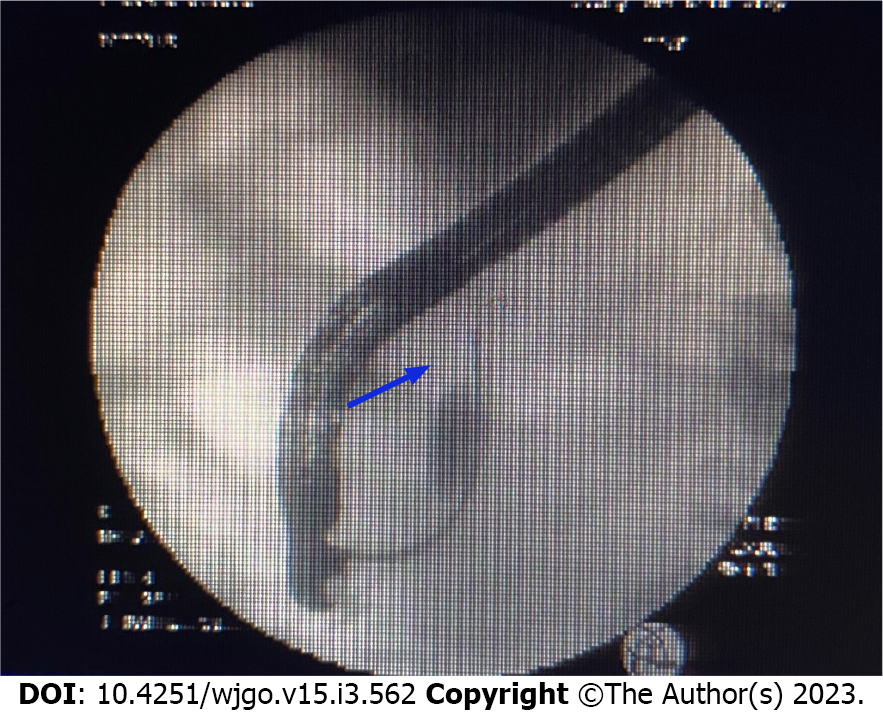
Figure 3 Endoscopic retrograde cholangiopancreatography was recommended for bile duct biopsy and a 7.
5 Fr × 7 cm bile duct plastic stent was implanted.
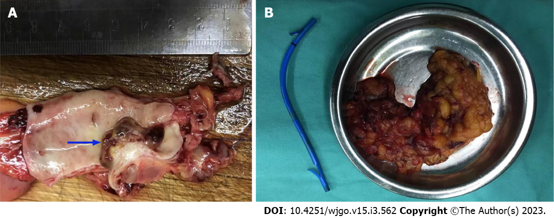
Figure 4 Intraoperative exploration revealed a tumor located in the middle and lower part of the common bile duct.
A: Cauliflower-like new organisms approximately 2 cm × 1.5 cm with a hard texture; B: Cholecystectomy, common bile duct resection, and choledochojejunostomy were performed.
Figure 5 Postoperative pathological examination revealed carcinosarcoma of the common bile duct with a tumor volume of 1.
0 cm × 0.6 cm × 0.6 cm. Cancerous tissue accounted for 40% and sarcoma for 60% of the tissue. A: Heterotypic glands can be seen in the mucosa of the bile duct, and infiltrating growth can be seen. Adenocarcinoma can be seen in the muscle wall of the bile duct; B: The muscle wall stroma of bile duct can be composed of heterotypic large cells, epithelial cancer nests and sarcoma-like components; C: Sarcoma-like area, large nuclear heterotypic cells in various forms and pathological mitotic image can be seen.
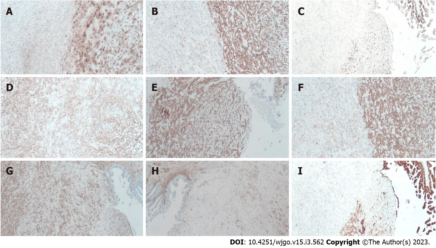
Figure 6 Immunohistochemical examination findings.
A: The tissue was positive for carbohydrate antigen 19-9; B: The tissue was positive for CK7; C: Ki-67 was approximately 40%; D: The tissue was positive for Calponin; E: The tissue was positive for CK19; F: The tissue was positive for CKpan; G: The tissue was positive for vimentin; H: The tissue was positive for S-100p; I: The tissue was negative for spinal muscular atrophy.
- Citation: Yao Y, Xiang HG, Jin L, Xu M, Mao SY. Carcinosarcoma of common bile duct: A case report. World J Gastrointest Oncol 2023; 15(3): 562-570
- URL: https://www.wjgnet.com/1948-5204/full/v15/i3/562.htm
- DOI: https://dx.doi.org/10.4251/wjgo.v15.i3.562


