Copyright
©The Author(s) 2023.
World J Gastrointest Oncol. Feb 15, 2023; 15(2): 286-302
Published online Feb 15, 2023. doi: 10.4251/wjgo.v15.i2.286
Published online Feb 15, 2023. doi: 10.4251/wjgo.v15.i2.286
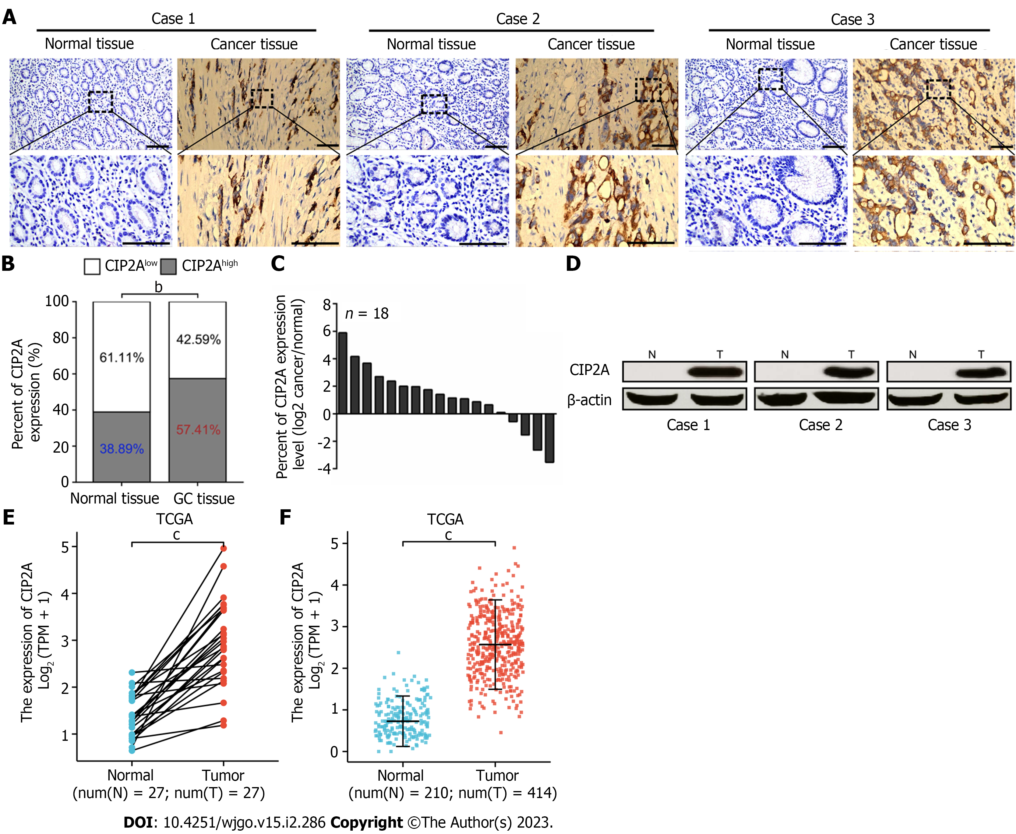
Figure 1 Cancerous inhibitor of protein phosphatase 2A was highly expressed in gastric cancer tissues.
A: Representative immunohistochemistry images of cancerous inhibitor of protein phosphatase 2A (CIP2A) in the adjacent normal gastric tissues and gastric cancer (GC) tissues (scale bar = 50 μm); B: The percentage of CIP2Ahigh expression was significantly higher in GC tissues than in adjacent normal gastric tissues; C: The mRNA expression levels of CIP2A were higher in 18 fresh frozen GC tissues than in matched adjacent normal gastric tissues; D: Western blot analysis showed that CIP2A expression level in tumor tissues (T) was significantly higher than that in adjacent normal tissues (N). Representative images were displayed in paired fresh surgical GC tissues; E and F: Based on data in The Cancer Genome Atlas (TCGA), including paired sample data and unpaired sample data (F), GC tissues had a higher CIP2A expression than adjacent normal gastric tissues. bP < 0.01. cP < 0.001.
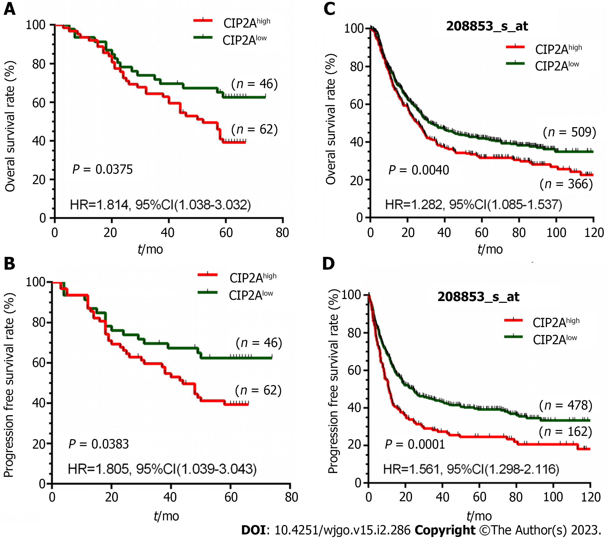
Figure 2 High expression of cancerous inhibitor of protein phosphatase 2A was associated with poor overall survival rate and progression-free survival rate in patients with gastric cancer.
A: Based on the Kaplan–Meier analysis, a high cancerous inhibitor of protein phosphatase 2A (CIP2A) expression was associated with overall survival (P = 0.0375) in 108 patients with gastric cancer; B: Based on the Kaplan–Meier analysis, a high CIP2A expression was associated with progression-free survival (P = 0.0383) in 108 patients with gastric cancer; C and D: According to the Kaplan–Meier analysis of data on gastric cancer from the 208853_s_at dataset, patients with CIP2Ahigh expression tumors had a significantly lower overall survival rate (P = 0.0040) (C) and progression-free survival rate (P < 0.0001) (D) than those with CIP2Alow expression tumors. CI: Confidence interval; HR: Hazard ratio.
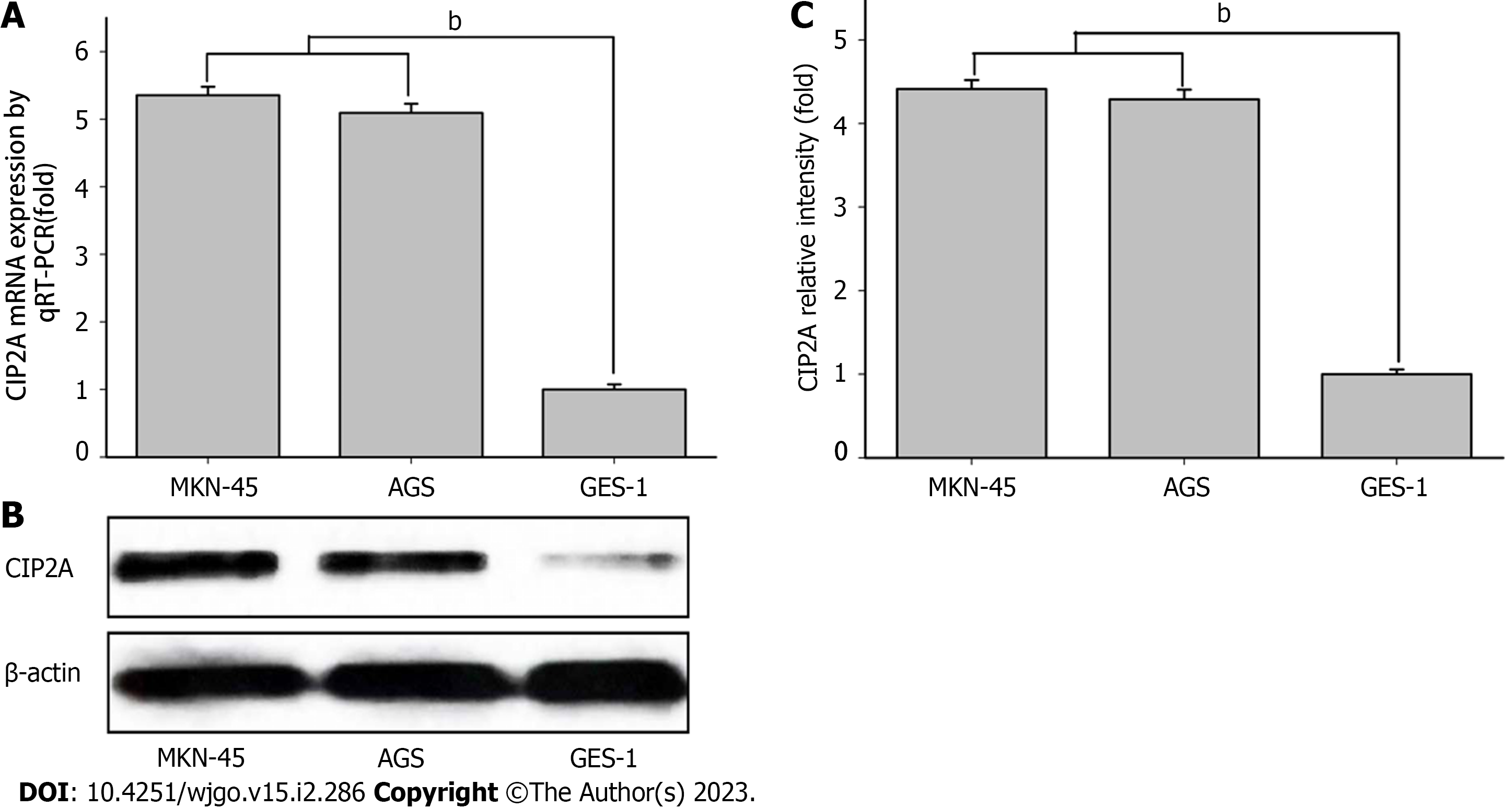
Figure 3 Expression of cancerous inhibitor of protein phosphatase 2A in human gastric cancer cells.
A: The mRNA expression of cancerous inhibitor of protein phosphatase 2A (CIP2A) in human gastric cancer cell lines (MKN-45 and AGS) and normal gastric epithelial cells (GES-1); B: The expression of cancerous inhibitor of protein phosphatase 2A was examined via immunoblotting; C: Immunoblotting was quantified using a spectrophotometer. The results consisted of three independent tests. Gastric cancer cell lines vs GES-1. bP < 0.01. RT: Reverse transcription.
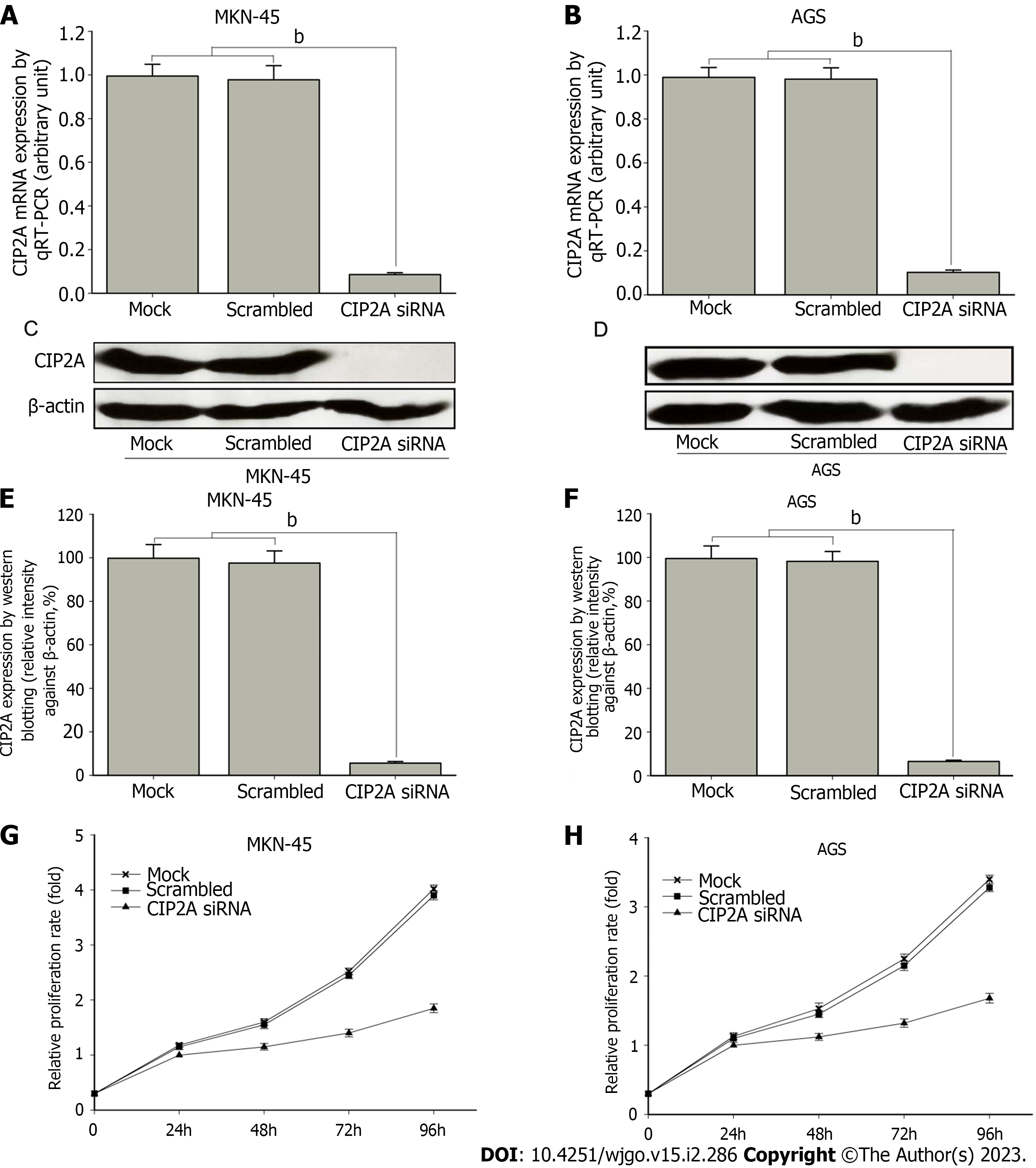
Figure 4 Small interference RNA targeting cancerous inhibitor of protein phosphatase 2A effectively downregulated expression and decreased cell proliferation in human gastric cancer cells.
MKN-45 and AGS cells were transfected with negative control small interference RNA (siRNA) or cancerous inhibitor of protein phosphatase 2A (CIP2A) siRNA for 48 h. A: CIP2A expression in MKN-45 cells was determined via real-time quantitative reverse transcription (RT) PCR; B: CIP2A expression in AGS cells was determined via real-time quantitative RT PCR; C and D: CIP2A expression was determined via immunoblotting; E and F: Data were quantified using a spectrophotometer. The CIP2A siRNA group vs the control siRNA group. G and H: The downregulation of CIP2A expression could decrease cell proliferation in MKN-45 and AGS cells based on the 3-(4,5-dimethylthiazol-2-yl)-2,5-diphenyltetrazolium bromide tetrazolium assay. Data showed a significant reduction in the proliferation of CIP2A siRNA-treated cells. The results were three independent tests. bP < 0.01.
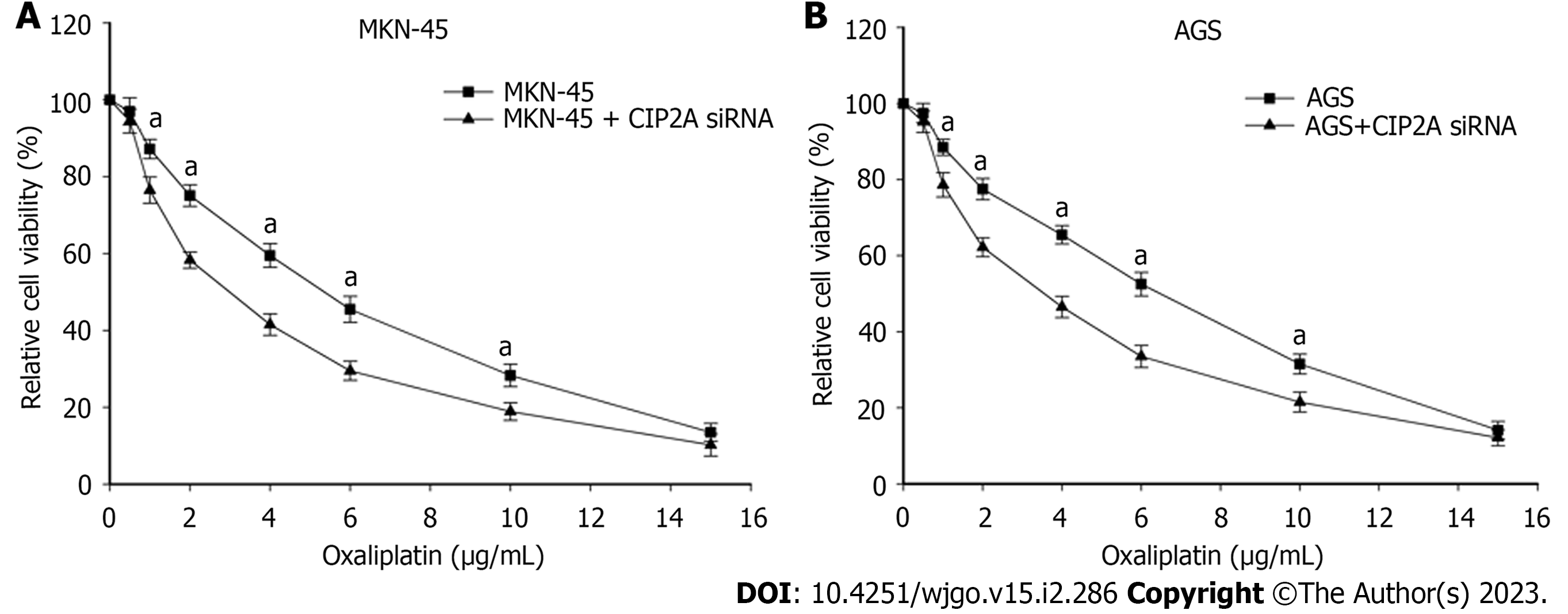
Figure 5 Downregulation of cancerous inhibitor of protein phosphatase 2A significantly increased sensitivity to oxaliplatin in human gastric cancer cells.
A: MKN-45 cells were transfected with cancerous inhibitor of protein phosphatase 2A (CIP2A) small interfering RNA (siRNA) for 48 h and were added with oxaliplatin at different concentrations for 24 h; B: AGS cells were transfected with cancerous inhibitor of protein phosphatase 2A siRNA for 48 h and were added with oxaliplatin at different concentrations for 24 h. The cell viability was examined using the 3-(4,5-dimethylthiazol-2-yl)-2,5-diphenyltetrazolium bromide tetrazolium assay. The results consisted of three independent tests. The siRNA group vs the control siRNA group. bP < 0.01.
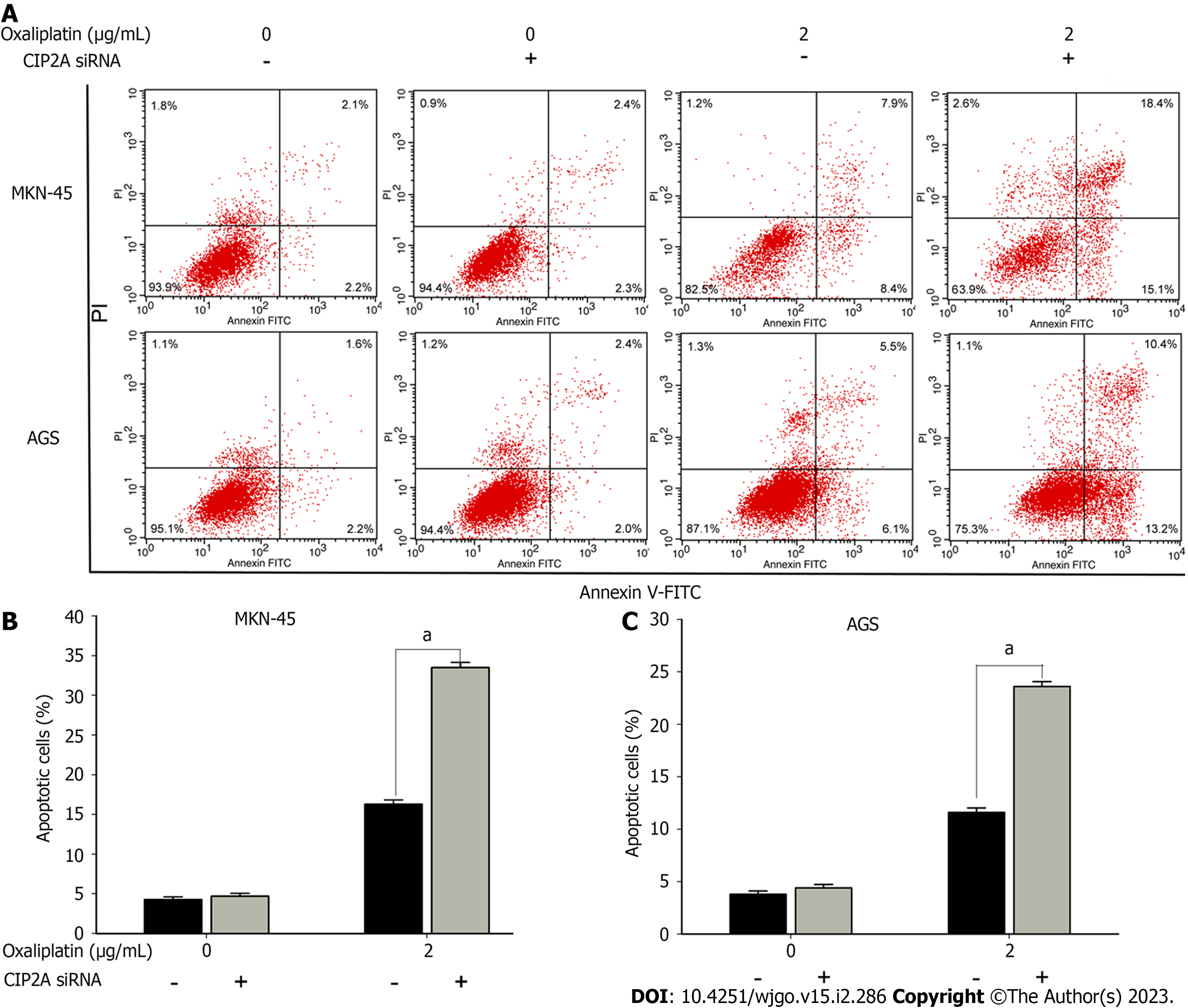
Figure 6 Downregulation of cancerous inhibitor of protein phosphatase 2A promoted oxaliplatin-related apoptosis in human gastric cancer cells.
Control small interference RNA (siRNA) and cancerous inhibitor of protein phosphatase 2A (CIP2A) siRNA-transfected human gastric cancer cells were exposed to oxaliplatin (2 μg/mL). A: At 24 h after treatment, apoptosis was examined via annexin V/propidium iodide staining and flow cytometry; B and C: The percentage of apoptotic cells was quantitatively presented in MKN-45 (B) and AGS (C) cells. The results consisted of three independent tests. The cancerous inhibitor of protein phosphatase 2A siRNA group vs the control siRNA group. aP < 0.05. V-FITC: V-fluorescein 5-isothiocyanate.
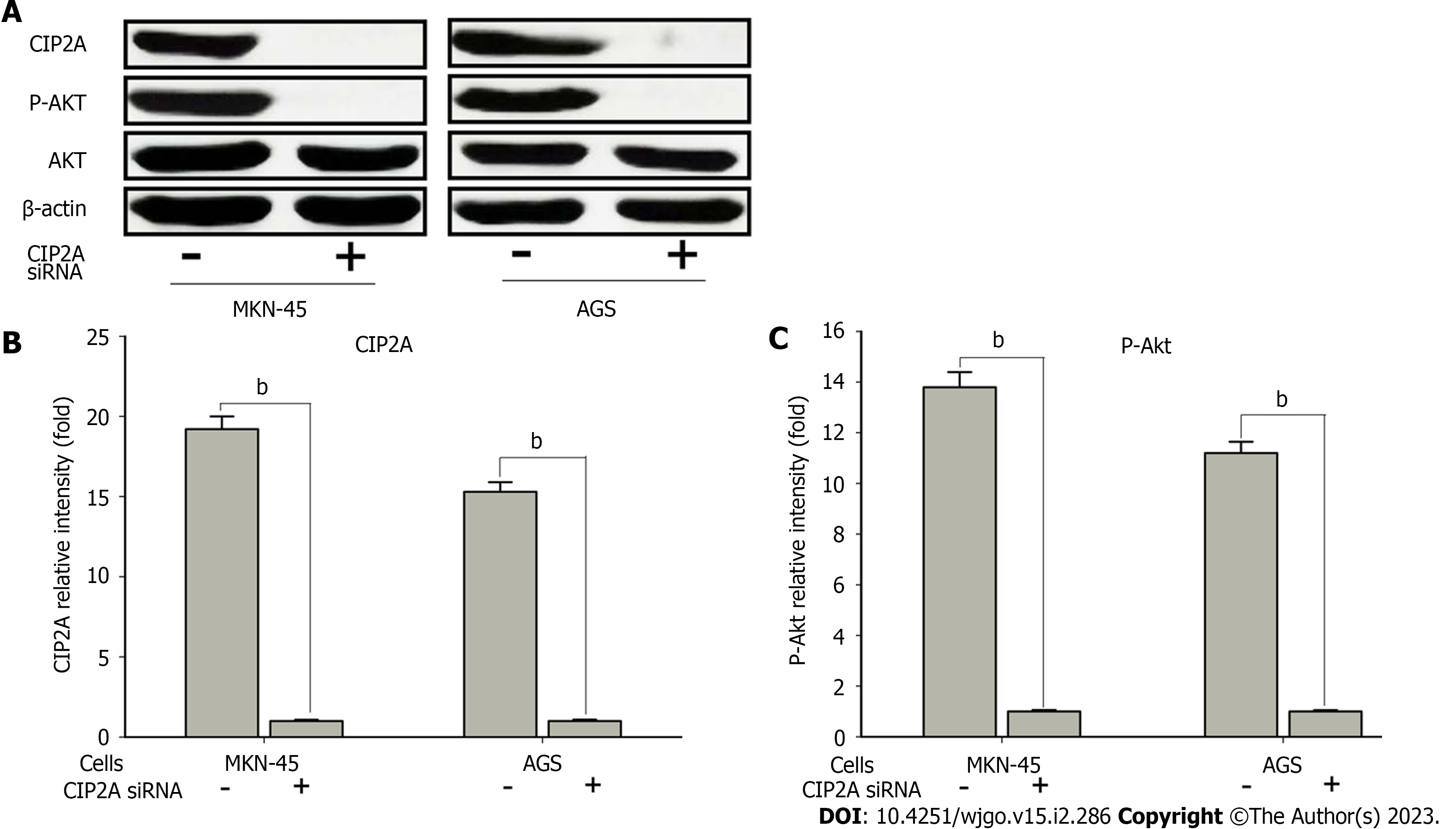
Figure 7 Cancerous inhibitor of protein phosphatase 2A affected protein kinase B phosphorylation in human gastric cancer cells.
MKN-45 and AGS cells were transfected with the control small interfering RNA (siRNA) or cancerous inhibitor of protein phosphatase 2A (CIP2A) siRNA for 48 h. A: Immunoblotting was used to analyze the indicated protein expression; B and C: The CIP2A (B) and phosphorylated protein kinase B (p-Akt) (C) protein levels were quantified using a spectrophotometer. The results consisted of three independent tests. bP < 0.01. Akt: Protein kinase B.
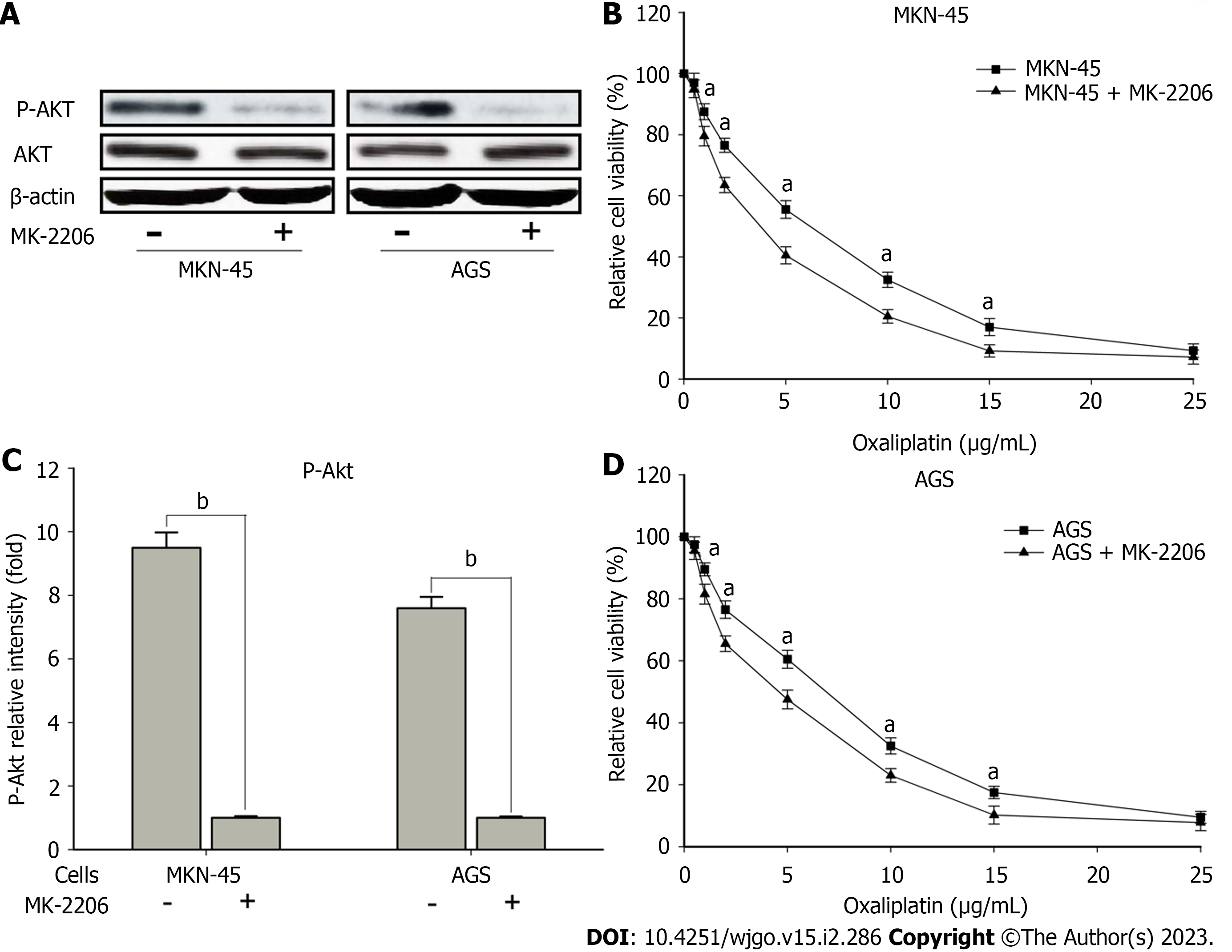
Figure 8 Chemical inhibition of protein kinase B signaling significantly increased sensitivity to oxaliplatin in human gastric cancer cells.
MKN-45 and AGS cells were added to inhibitor of protein kinase B (MK-2206; 20 μM) for 2 h. A: Immunoblotting was performed to evaluate the corresponding protein expression; B: The phosphorylated protein kinase B (P-Akt) protein levels were quantified via densitometry; C and D: The pretreated cells were exposed to oxaliplatin at different concentrations for 24 h, and the viability was examined using the 3-(4,5-dimethylthiazol-2-yl)-2,5-diphenyltetrazolium bromide tetrazolium assay. The results consisted of three independent tests. The MK-2206-treated group vs the control group. aP < 0.05. bP < 0.01. Akt: Protein kinase B.
- Citation: Zhao YX, Ma LB, Yang Z, Wang F, Wang HY, Dang JY. Cancerous inhibitor of protein phosphatase 2A enhances chemoresistance of gastric cancer cells to oxaliplatin. World J Gastrointest Oncol 2023; 15(2): 286-302
- URL: https://www.wjgnet.com/1948-5204/full/v15/i2/286.htm
- DOI: https://dx.doi.org/10.4251/wjgo.v15.i2.286


