Copyright
©The Author(s) 2023.
World J Gastrointest Oncol. Nov 15, 2023; 15(11): 1891-1899
Published online Nov 15, 2023. doi: 10.4251/wjgo.v15.i11.1891
Published online Nov 15, 2023. doi: 10.4251/wjgo.v15.i11.1891
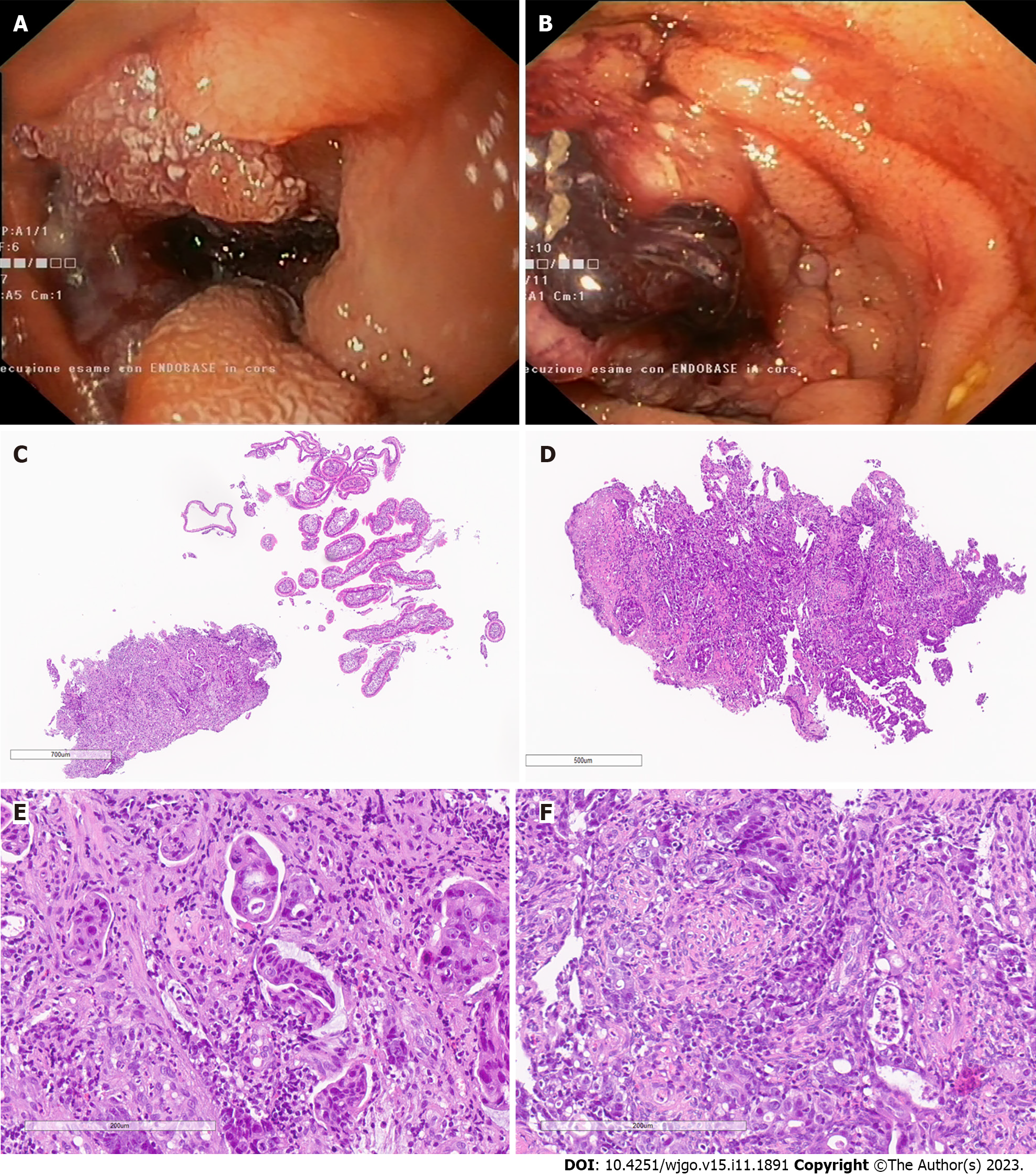
Figure 1 Endoscopic and histopathological features of the two cases of small bowel cancer identified.
A: Upper push-enteroscopy in the first patient showed a vegetate-ulcerative lesion located 100-105 cm from the incisors, involving at least three-quarters of the luminal circumference, that causes partial stenosis of the lumen that could be passed through, anyway. There was a small clot in the distal portion, but the lesion was not actively bleeding. Multiple biopsies were taken, and the diagnosis was adenocarcinoma of the distal duodenum; B: Upper push-enteroscopy in the second patient showed an ulcerative lesion located 120-130 cm from the incisors, actively bleeding, involving at least two-thirds of the luminal circumference, with a clot in the mid portion. Multiple biopsies were taken and after clot removal, hemospray was applied to the tumor surface and complete hemostasis was obtained. Tattooing was then performed. The diagnosis was adenocarcinoma of the proximal jejunum; C: Hematoxylin-eosin staining in the first patient, 5 ×. Low magnification reveals two bioptic fragments. The first, on the right, of normal small intestine and the second, on the left, of fibrous/granulation tissue, show variably sized glands and desmoplasia, indicative of adenocarcinoma; D: Hematoxylin-eosin staining in the second patient, 5 ×. Low magnification reveals a single bioptic fragment of fibrous tissue showing variably sized glands with moderate/severe atypia (loss of normal glandular architecture) and desmoplasia, indicative of adenocarcinoma; E: Hematoxylin-eosin staining in the first patient, 20 ×. On higher magnification, solid nests and sheets and few and small poorly-formed glands contain enlarged, hyperchromatic cells with loss of mucin, nuclei with prominent eosinophilic nucleoli and irregular nuclear membranes in a desmoplastic stroma, diagnostic of invasive adenocarcinoma; F: Hematoxylin-eosin staining in the second patient, 20 ×. High magnification shows marked cytological atypia (the cells are hyperchromatic with loss of mucin and contain nuclei with prominent eosinophilic nucleoli and irregular nuclear membranes) forming marked atypical glands with intraluminal apoptotic and inflammatory debris in a desmoplastic stroma, suggestive of invasive adenocarcinoma.
- Citation: Sanchez-Mete L, Mosciatti L, Casadio M, Vittori L, Martayan A, Stigliano V. MUTYH-associated polyposis: Is it time to change upper gastrointestinal surveillance? A single-center case series and a literature overview. World J Gastrointest Oncol 2023; 15(11): 1891-1899
- URL: https://www.wjgnet.com/1948-5204/full/v15/i11/1891.htm
- DOI: https://dx.doi.org/10.4251/wjgo.v15.i11.1891


