Copyright
©The Author(s) 2023.
World J Gastrointest Oncol. Jan 15, 2023; 15(1): 186-194
Published online Jan 15, 2023. doi: 10.4251/wjgo.v15.i1.186
Published online Jan 15, 2023. doi: 10.4251/wjgo.v15.i1.186
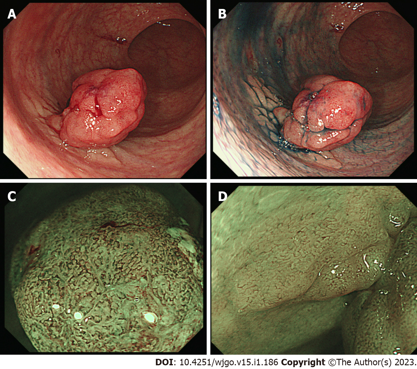
Figure 1 Endoscopic findings of the rectal tumor.
A: A remarkable protrusion (Is) with slight bleeding is observed in the rectum; B: Chromoendoscopy enhances a flat elevated lesion (IIa) which is located at the base of the protrusion lesion; C: Magnified endoscopy with narrow-band imaging reveals an intense irregular micro-vascular pattern indicating the existence of carcinoma in the Is lesion; D: Magnified endoscopy shows faint vascular pattern on the IIa lesion.
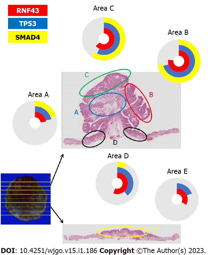
Figure 2 A comparison between the genetic mutations and the histopathological characteristics of the tumor.
Mucinous and signet-cell carcinoma (area A) and tubular adenocarcinoma (area B, C) shows the same mutational frequency in RNF43, TP53, and SMAD4. The adenoma (area D, E) shows a higher frequency for RNF 43 than for TP53.
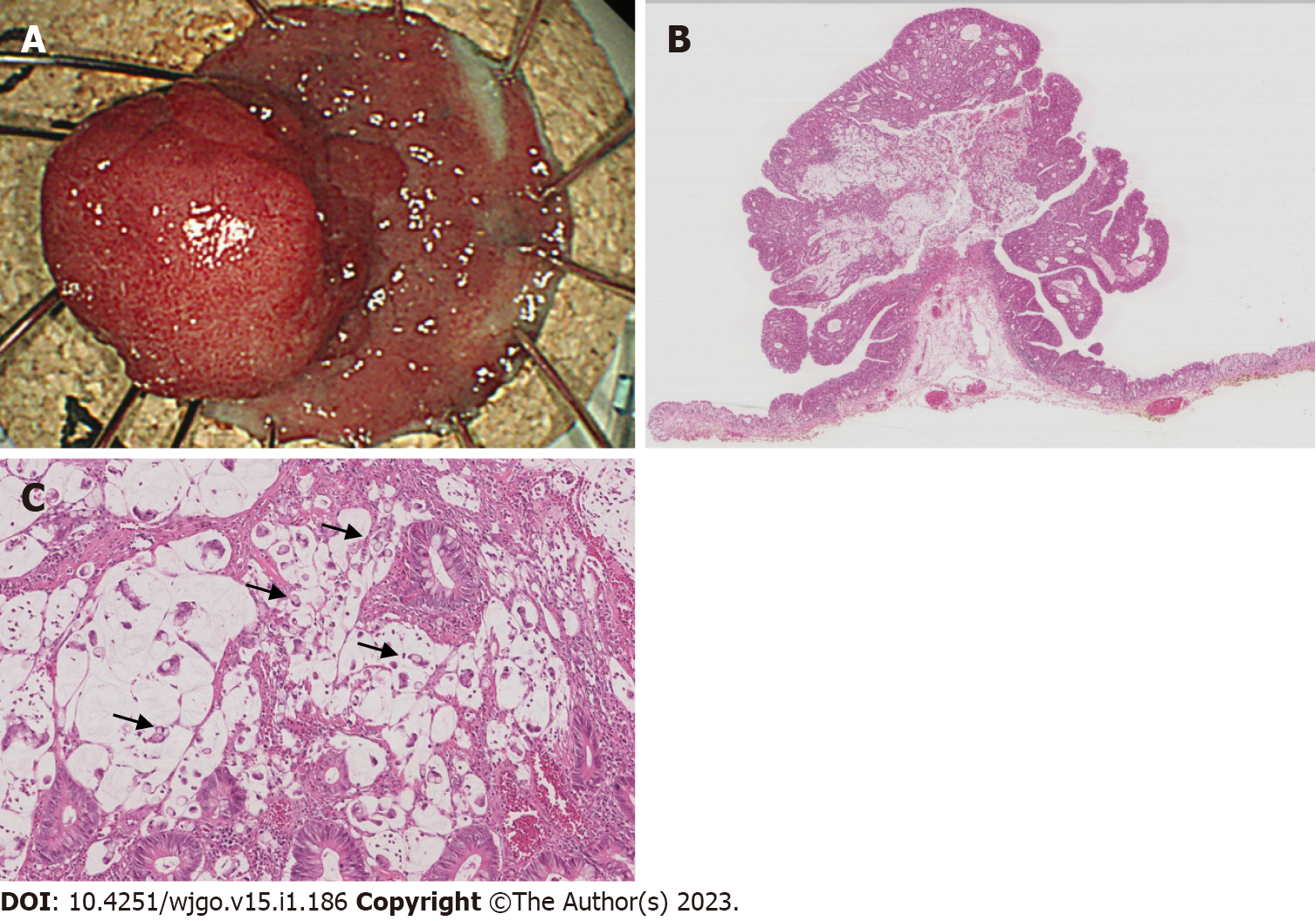
Figure 3 A histopathological examination of the endoscopically resected specimen.
A: A protruded polyp and flat elevation are removed by endoscopic submucosal dissection. The tumor margin is surrounded by normal epithelia, indicating R0 resection; B: Adenocarcinoma composed of mucinous and tubular carcinoma with an adenoma component; C: Signet ring cell carcinoma is observed in mucinous lake (arrows).
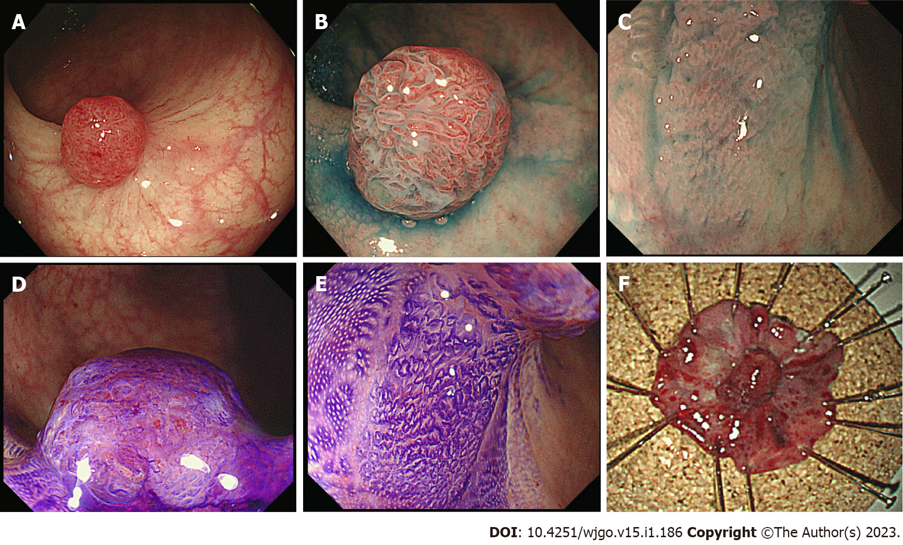
Figure 4 Endoscopic findings of the recurrent tumor.
A: A protrusion tumor (Is) with redness is observed on the scar after endoscopic resection; B: Chromoendoscopy shows an elongated tubular surface tumor with a hypervascular pattern on the Is lesion; C: The flat elevated lesion (IIa) located at the base of Is tumor indicates dilated crypts; D: Crystal violet staining indicates irregular structured pits on the Is; E: Magnified endoscopy shows round crypts with a sessile pit pattern on the IIa lesion; F: Macroscopic view of the specimen resected by re-endoscopic submucosal dissection. The removed Is+IIa lesion is surrounded by normal mucosa.
- Citation: Murakami Y, Tanabe H, Ono Y, Sugiyama Y, Kobayashi Y, Kunogi T, Sasaki T, Takahashi K, Ando K, Ueno N, Kashima S, Yuzawa S, Moriichi K, Mizukami Y, Fujiya M, Okumura T. Local recurrence after successful endoscopic submucosal dissection for rectal mucinous mucosal adenocarcinoma: A case report. World J Gastrointest Oncol 2023; 15(1): 186-194
- URL: https://www.wjgnet.com/1948-5204/full/v15/i1/186.htm
- DOI: https://dx.doi.org/10.4251/wjgo.v15.i1.186


