Copyright
©The Author(s) 2022.
World J Gastrointest Oncol. Sep 15, 2022; 14(9): 1833-1843
Published online Sep 15, 2022. doi: 10.4251/wjgo.v14.i9.1833
Published online Sep 15, 2022. doi: 10.4251/wjgo.v14.i9.1833
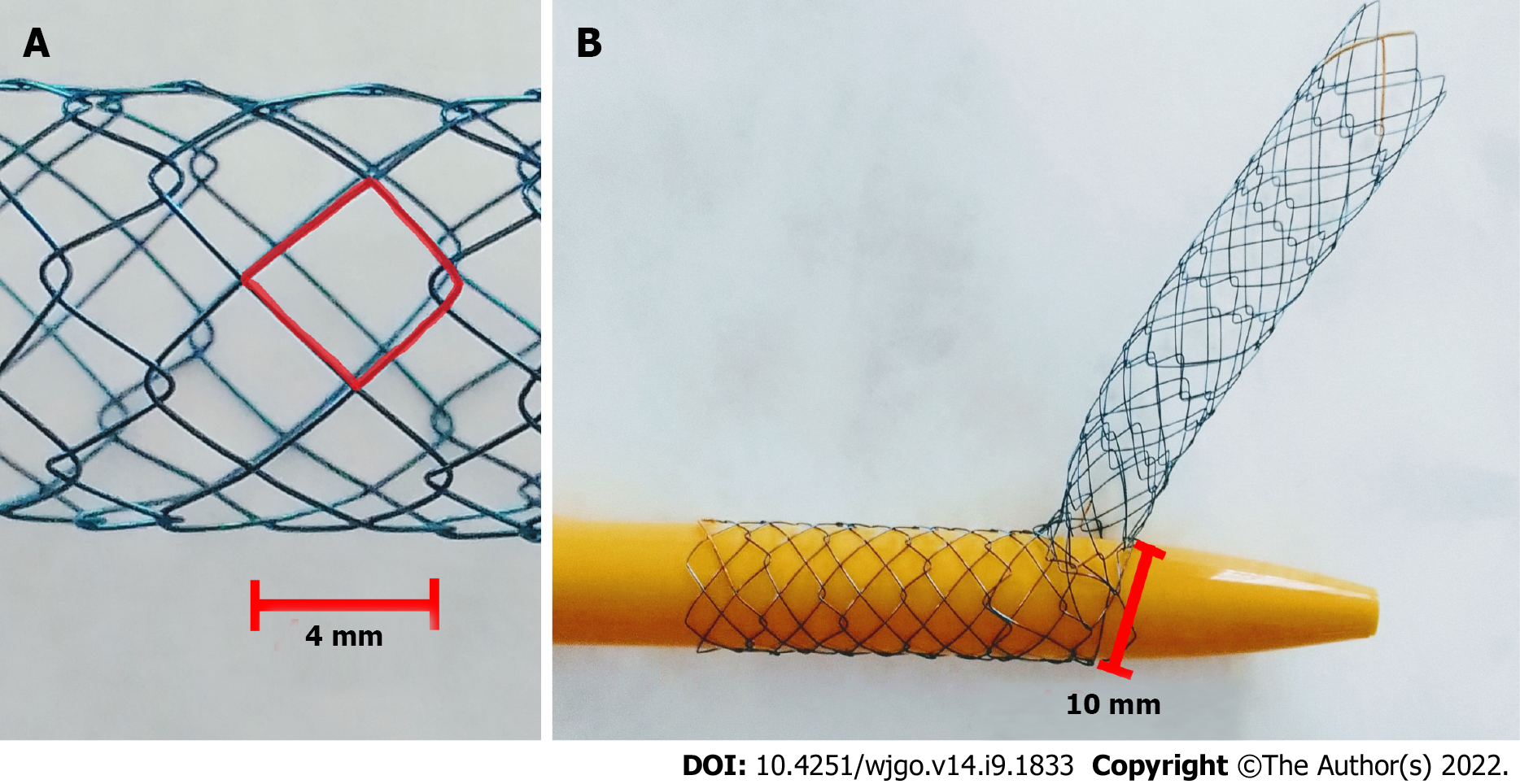
Figure 1 The Hilzo Biliary Moving Cell Stent.
A: The Hilzo Biliary Moving Cell Stent developed with small cell size (4 mm), with radiopaque markers at each end and two X-shape markers in the midsection; B: Each cell can expand from 4 mm to 10 mm to allows easier passage of the second stent through the cell.
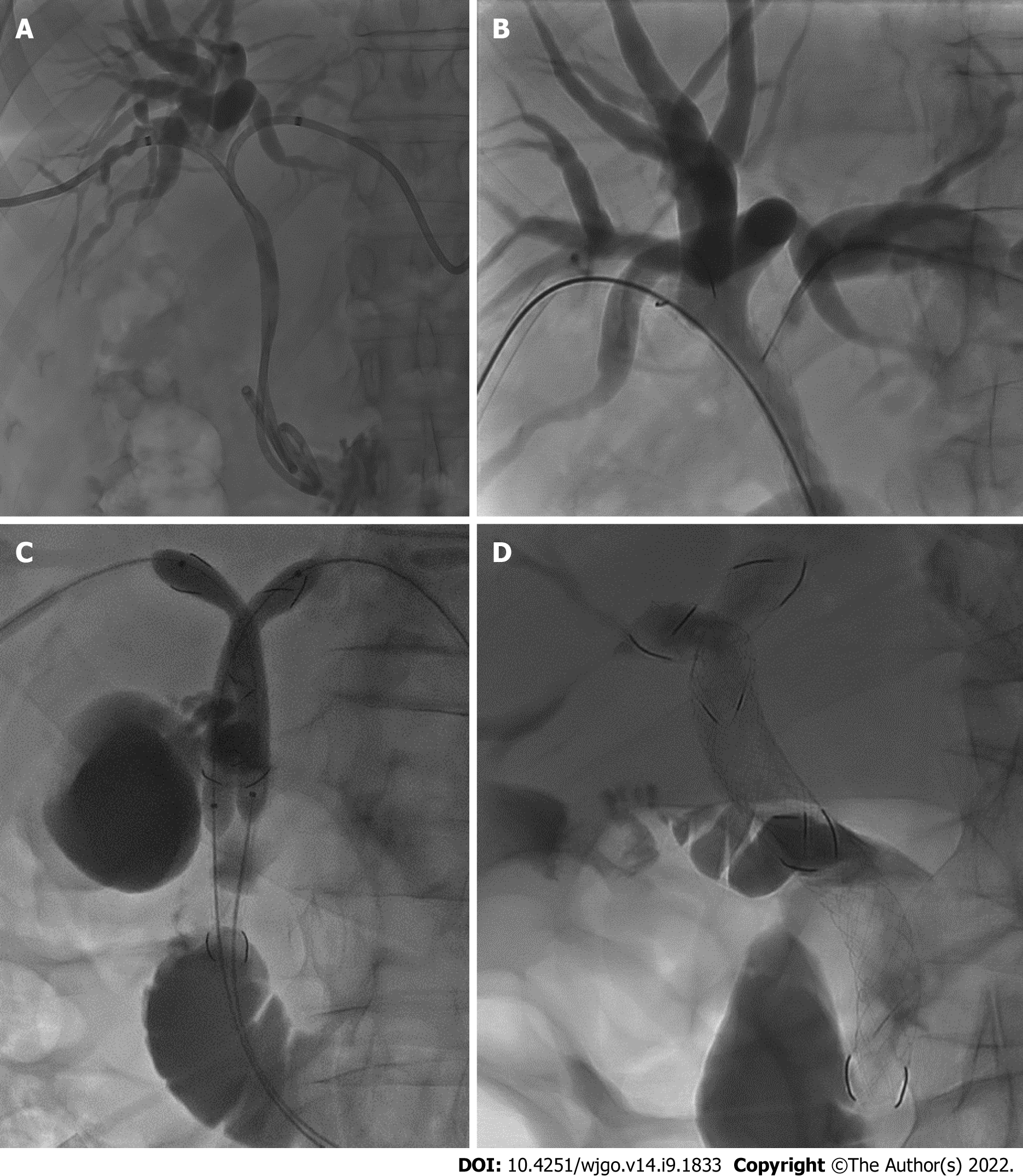
Figure 2 Percutaneous transhepatic cholangiography.
A: Percutaneous transhepatic cholangiography (PTC) showing hilar biliary obstructions with two bilateral bilateral 8.5-Fr drainage catheters; B: A hydrofilic guidewire (0.035 in.; Terumo Corporation, Tokyo, Japan) was inserted through a mesh of the Moving Cell Stent (MCS); C: PTC showing a kissing baloon dilatation over the stiff guidewires inside MCS placed using sten-in-stent technique; D: PTC showing the appropriate stents placement with the apex of the longest stent lies in the duodenum, while the apex of the shorter stent ends inside the first.
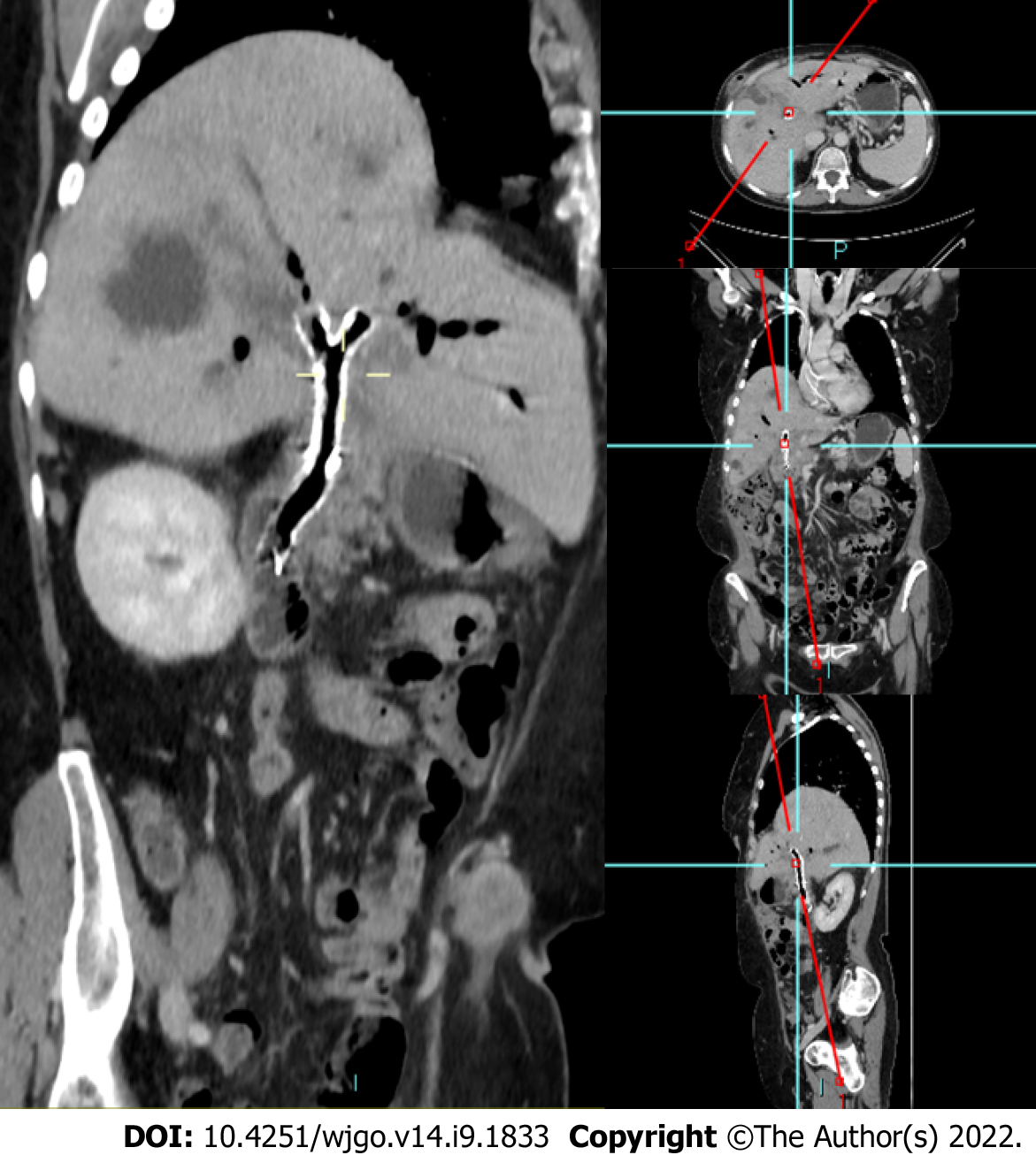
Figure 3 Three-months follow-up contrast-enhanced computed tomography.
Sagittal oblique MPR showing two Y-shape Moving Cell Stent placed at the hilar bifurcation biliary with no intrahepatic biliary dilatation.
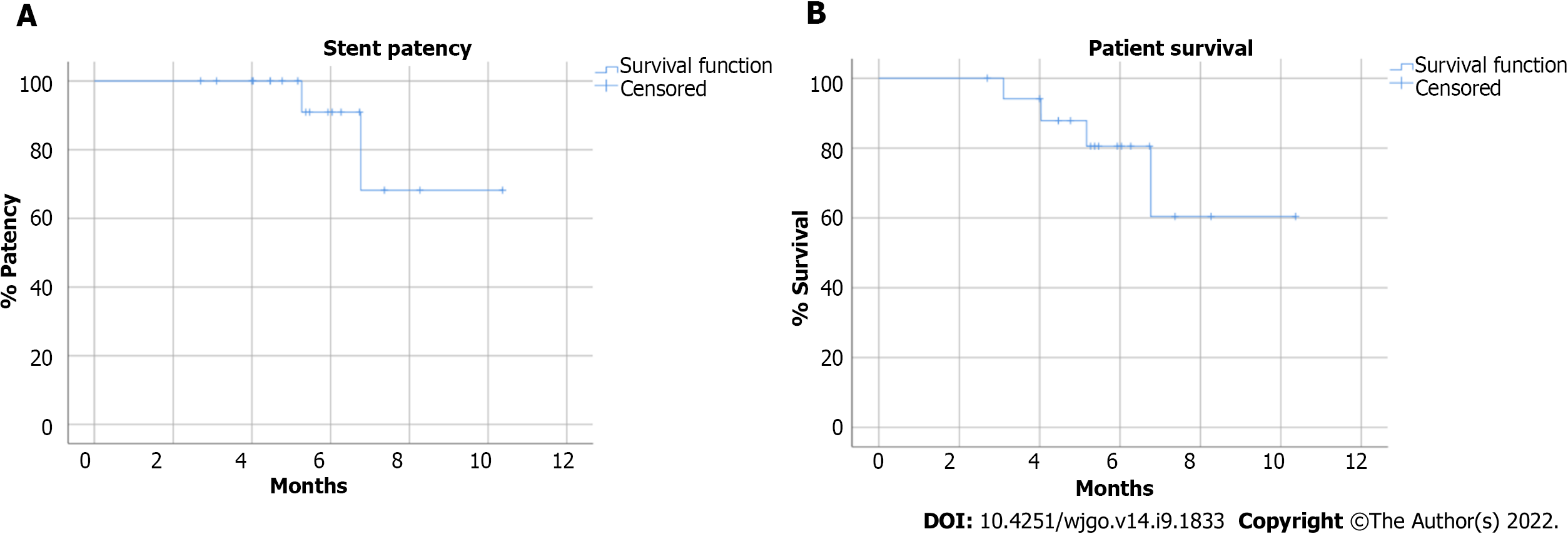
Figure 4 Kaplan-Meier analysis.
A: The estimated stent patency; B: Overall patient survival.
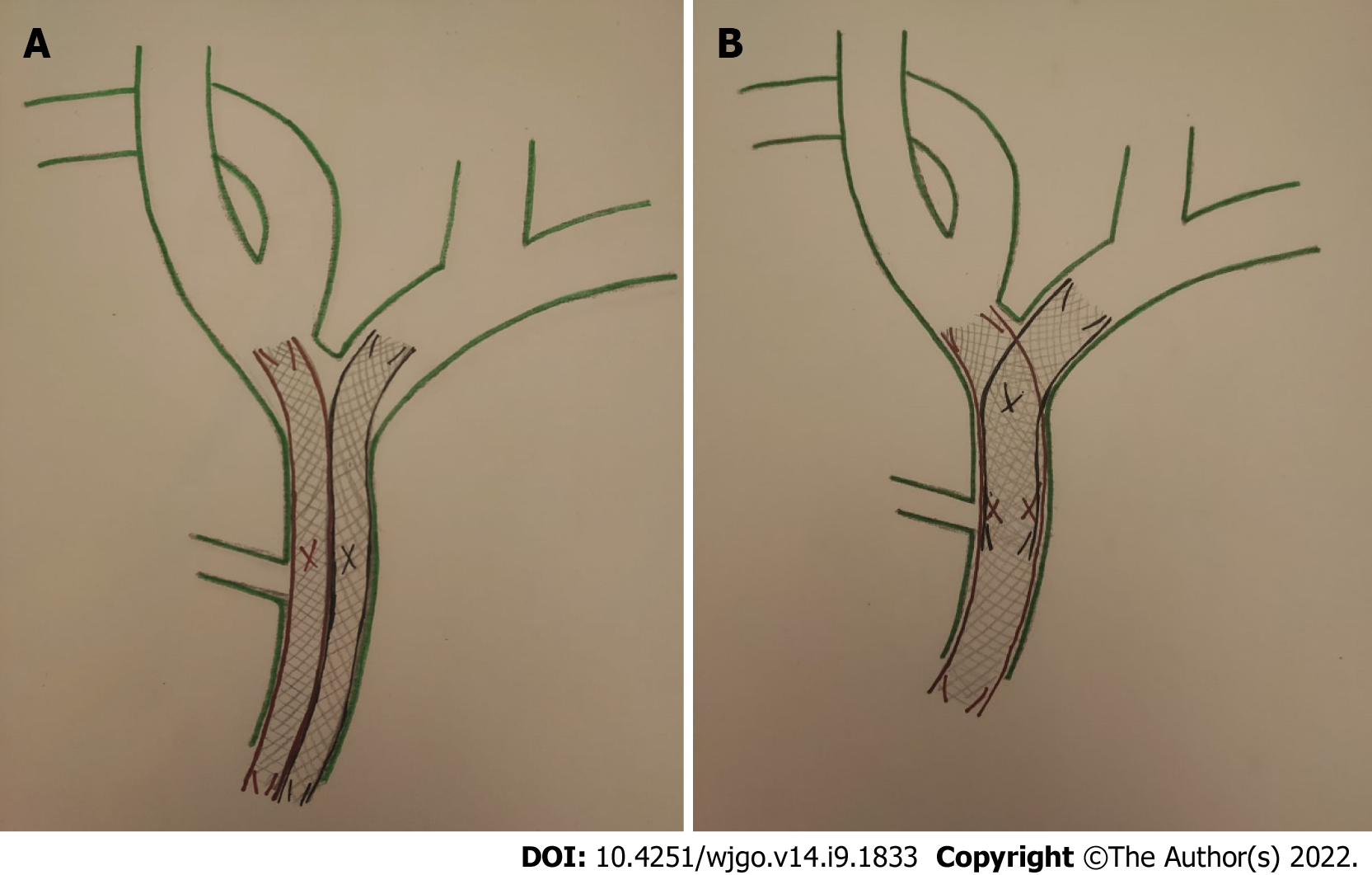
Figure 5 Bilateral self-expandable metallic stent placement can be achieved with side-by-side or stent-in-stent techniques.
A: Stent-by-stent technique: Two parallel and close self-expandable metallic stent (SEMS) at and below the hepatic confluence to drain the bile duct of both hepatic lobes; B: Stent-in-stent technique: Bilateral SEMS placed in a Y-configuration, in which a second stent across through the mesh of the first stent.
- Citation: Cortese F, Acquafredda F, Mardighian A, Zurlo MT, Ferraro V, Memeo R, Spiliopoulos S, Inchingolo R. Percutaneous insertion of a novel dedicated metal stent to treat malignant hilar biliary obstruction. World J Gastrointest Oncol 2022; 14(9): 1833-1843
- URL: https://www.wjgnet.com/1948-5204/full/v14/i9/1833.htm
- DOI: https://dx.doi.org/10.4251/wjgo.v14.i9.1833


