Copyright
©The Author(s) 2022.
World J Gastrointest Oncol. Aug 15, 2022; 14(8): 1585-1593
Published online Aug 15, 2022. doi: 10.4251/wjgo.v14.i8.1585
Published online Aug 15, 2022. doi: 10.4251/wjgo.v14.i8.1585
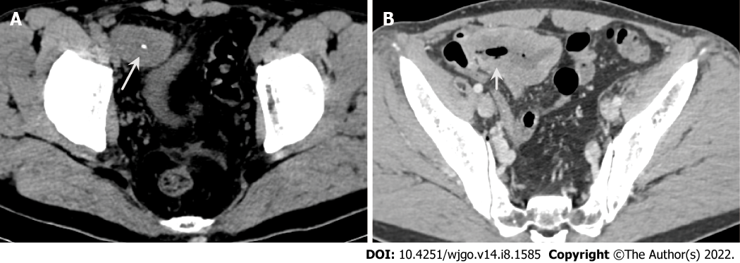
Figure 1 Abdominal computed tomography.
A: Axial computed tomography (CT) image shows a heterogenetic mass with calcification (white arrows); B: Contrast-enhanced CT shows mild heterogenetic enhancement and communication with the small intestinal lumen (short white arrows).
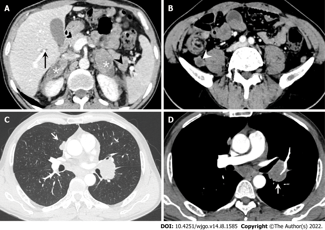
Figure 2 Abdominal and chest computed tomography.
A: Multiple metastatic lesions are observed on the bilateral adrenal gland (*), liver (black arrow) and pancreas (black arrowhead); B: Several enlarged lymph nodes (white arrowhead) are seen on the retroperitoneal area; C: A pulmonary metastatic nodule (short white arrow) is seen in lung windows; D: Several enlarged lymph nodes (short white arrow) are shown on the mediastinum area in contrast-enhanced computed tomography.
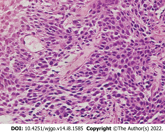
Figure 3 Pathologic findings.
Histopathology of the small intestinal tumor is composed of heteromorphic cells, and distributed in the shape of sheet or nest with round or oval cells and visible nucleoli. (Original magnification 400 ×; hematoxylin-eosin stains).
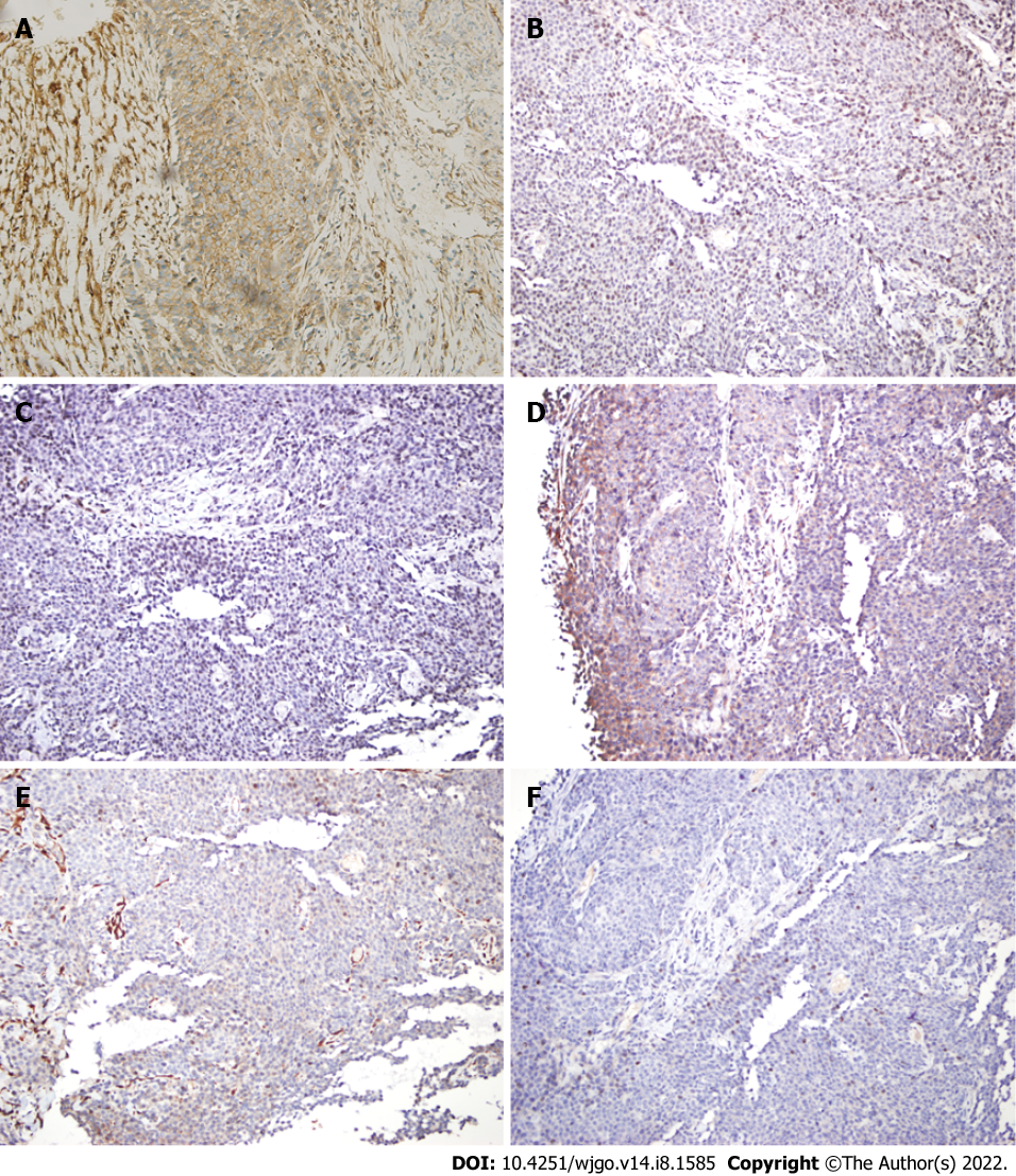
Figure 4 Immunohistochemistry findings.
A: Strong positive staining for CD99 (original magnification 200 ×); B: Positive staining for NKX2.2 original magnification 200 ×); C: Positive staining for FLI-1 (original magnification 200 ×); D: Positive staining for Syn (original magnification 200 ×); E: Negative immunoreactivity for CK (original magnification 200 ×); F: Negative immunoreactivity for CgA (original magnification 200 ×).
- Citation: Guo AW, Liu YS, Li H, Yuan Y, Li SX. Ewing sarcoma of the ileum with wide multiorgan metastases: A case report and review of literature. World J Gastrointest Oncol 2022; 14(8): 1585-1593
- URL: https://www.wjgnet.com/1948-5204/full/v14/i8/1585.htm
- DOI: https://dx.doi.org/10.4251/wjgo.v14.i8.1585


